
1. Guideline development and scope of the guideline
1.1 Guiding principles
The development of this guideline was guided by the following principles:
• Equity: Ensuring access to essential care for all women in Sri Lanka, regardless of socioeconomic status.
• Feasibility: Prioritizing recommendations that can be realistically implemented with available resources.
• Cost-Effectiveness: Advocating for interventions that provide the greatest health benefit for the investment.
• Simplicity: Providing clear, unambiguous recommendations and algorithms to minimize complexity at the point of care.
• Evidence-based practice: Ensuring that all recommendations are consistent with the best available international and local evidence, while adapting them to the Sri Lankan context.
1.2 Methodology of guideline development
1.2.1 Guideline committee
The guideline committee comprised a multidisciplinary team: guideline committee of the Sri Lanka College of obstetricians and gynaecologists (SLCOG) and Sri Lanka College of Haematologist (SLCH).
1.2.2 Literature search and synthesis
A systematic search for evidence was conducted in electronic databases: PubMed, Cochrane Library and cited reference search. The search focused on guidelines, systematic reviews, randomized controlled trials, observational studies and local publications.
1.2.3 Formulation of recommendations
Recommendations were formulated during consensus meetings of the guideline committee. Evidence was categorised according to the scope areas and presented to guideline committee that was analysed by the guideline committee considering following principles: balance of benefits and harms, the quality of evidence, cultural acceptance, and resource implications.
1.2.4 Peer review and approval
The draft guideline was subjected to external review by the consultant obstetricians and gynaecologists (SLCOG) and consultant haematologists (SLCH) who were not part of the guideline committee. Their feedback was collated, reviewed and incorporated in the final version of the guideline by the guideline committee.
1.2.5 Plan for update
This guideline is scheduled for a full review in 2028 or sooner if new, practice-changing evidence emerges. The joint guideline committee of SLCOG and SLCH will monitor for new evidence.
Scope of the guideline
The purpose of this guideline is to explore the epidemiology, risk factors, diagnostic challenges, and management strategies of thromboembolism in obstetrics and gynaecology within the Sri Lankan context.
By examining available data and identifying gaps in healthcare delivery, this aims to highlight the significance of thromboembolism as a preventable cause of maternal morbidity and mortality in Sri Lanka.
Also, thromboembolism preventive strategies are implemented in a limited manner among gynaecological patients in Sri Lanka. This gap in practice should be addressed through the development and dissemination of appropriate guidelines and increased awareness among healthcare professionals.
2. Introduction and background epidemiology of thromboembolism in pregnancy and gynaecology
2.1 Thromboembolism in pregnancy
Thromboembolism, including deep vein thrombosis (DVT) and pulmonary embolism (PE), is a leading cause of maternal mortality worldwide, accounting for approximately 10% of maternal deaths in high-income countries (1). The risk of venous thromboembolism (VTE) during pregnancy is four to five times higher than in non-pregnant women, with an incidence of 1-2 cases per 1,000 pregnancies (2). In Sri Lanka, the maternal mortality ratio (MMR) is 25 per 100,000 live births in 2023 (3). Among the causes of maternal mortality, thromboembolism remains a critical issue, particularly in high-risk pregnancies. Although national data on thromboembolism in pregnancy remain limited, annual reports from the Family Health Bureau indicate an incidence of three per 10,000 deliveries per year between 2018 and 2022 (3). This emphasises the critical need for enhanced awareness and the establishment of systematic data collection mechanisms to improve monitoring and management. (6)
A systematic review done in 2017 stated that venous thromboembolism in Asian population is lower than that of in western population. Incidence estimates in Asia were approximately 15 to 20% of the levels recorded in western countries but have increased over time (4).
Risk factors for thromboembolism in pregnancy include advanced maternal age, obesity, multiparity, caesarean delivery, and a personal or family history of VTE (5). In Sri Lanka, cultural practices such as prolonged bed rest during pregnancy and the rising prevalence of obesity, diabetes in pregnancy and IVF pregnancies further exacerbate these risks.
The caesarean section rate in Sri Lanka, which reached 43.1% in 2022 (3), significantly increases the risk of VTE, underscoring the need for tailored thromboprophylaxis protocols. Diagnosing thromboembolism in pregnancy is challenging due to overlapping symptoms with normal physiological changes, such as lower extremity oedema and dyspnoea. However, cost, availability barriers and lack of standardised national guidelines hinder effective care.
Thromboembolism in gynaecology
In addition to the above, venous thromboembolism (VTE), including deep vein thrombosis (DVT) and pulmonary embolism (PE), is a major postoperative complication in gynaecological patients. Risk is particularly high following major gynaecological surgeries, malignancies, and assisted reproductive techniques (ART). Without thromboprophylaxis, the incidence of confirmed deep vein thrombosis (DVT) in major abdominal surgery ranges from 17 to 40%. Despite continued efforts to reduce its incidence, postoperative VTE remains the second most common perioperative complication and the third most common cause of mortality (6).
Maternal mortality and morbidity figures in Sri Lanka
Sri Lanka has made significant progress in reducing maternal mortality over the years, achieving one of the lowest maternal mortality ratios (MMR) in South Asia at 25 maternal deaths per 100,000 live births in 2023. (Figure 1)
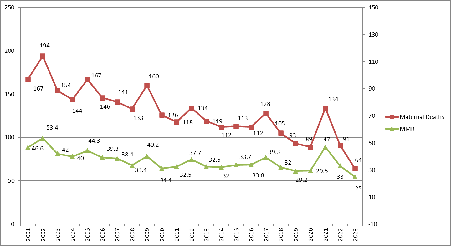
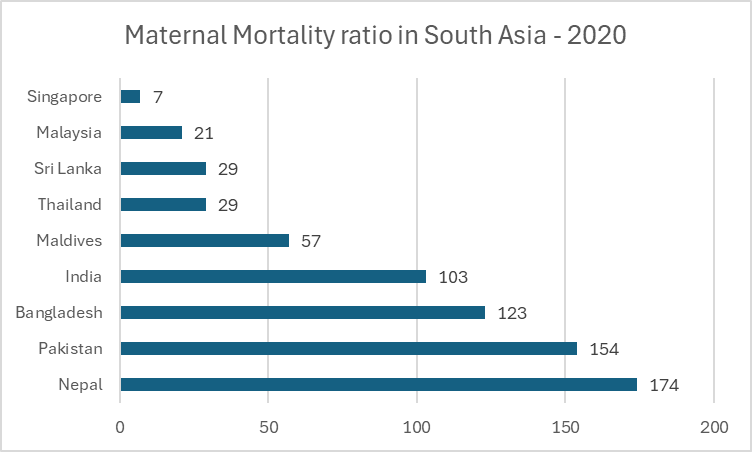
Sri Lanka's maternal mortality figures are considerably lower than those of neighbouring South Asian countries (Figure 2).
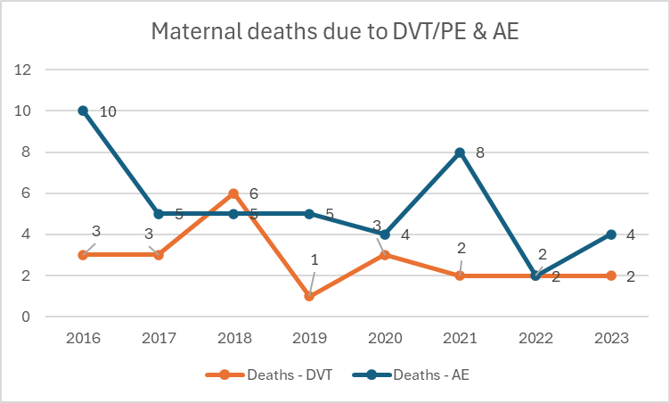
Though amniotic fluid embolism (AFE) remains an inherently non-preventable cause of maternal death, thromboembolism is a clearly preventable entity. National maternal mortality trends consistently show that thromboembolic deaths persist despite being avoidable, underscoring the need for strengthened, systematic preventive strategies (Table 1, Figure 3).
Table 1: Comparison of mortality due to VTE & AFE over the years in Sri Lanka
| Year | Number of deaths due to VTE | Number of deaths due to AFE |
|---|---|---|
| 2016 | 3 | 10 |
| 2017 | 3 | 5 |
| 2018 | 6 | 5 |
| 2019 | 1 | 5 |
| 2020 | 3 | 4 |
| 2021 | 2 | 8 |
| 2022 | 2 | 2 |
| 2023 | 2 | 4 |
References
1. Say L, Chou D, Gemmill A, Tunçalp Ö, Moller A-B, Daniels J, et al. Global causes of maternal death: a WHO systematic analysis. Lancet Glob Health. 2014;2(6):e323–33.
2. Heit JA, Kobbervig CE, James AH, Petterson TM, Bailey KR, Melton LJ. Trends in the incidence of venous thromboembolism during pregnancy or postpartum: a 30-year population-based study. Ann Intern Med. 2005;143(10):697–706.
3. Family Health Bureau. Annual Report on Maternal and Child Health. Colombo: Ministry of Health, Sri Lanka; 2022.
4. Scheres LJJ, Lijfering WM, Middeldorp S. Pregnancy, thrombophilia, and the risk of a first venous thrombosis: systematic review and meta-analysis. Thromb Haemost. 2017. Available from: [https://gynaecolo.thieme-connect.com/products/ejournals/pdf/10.1160/TH17-02-0134.pdf](https://gynaecolo.thieme-connect.com/products/ejournals/pdf/10.1160/TH17-02-0134.pdf)
5. National Guideline Alliance (UK). Risk factors for venous thromboembolism in pregnancy: antenatal care: evidence review N. London: National Institute for Health and Care Excellence (NICE); 2021 Aug. (NICE Guideline No. 201).
6. Rahn DD, Mamik MM, Sanses TVD, Matteson KA, Aschkenazi SO, Washington BB, et al. Venous thromboembolism prophylaxis in gynecologic surgery: a systematic review. Obstetrics & Gynecology. 2011;118(5):1111–25.
3. Risk assessment for VTE in pregnancy
3.1 Categorisation of risk factors
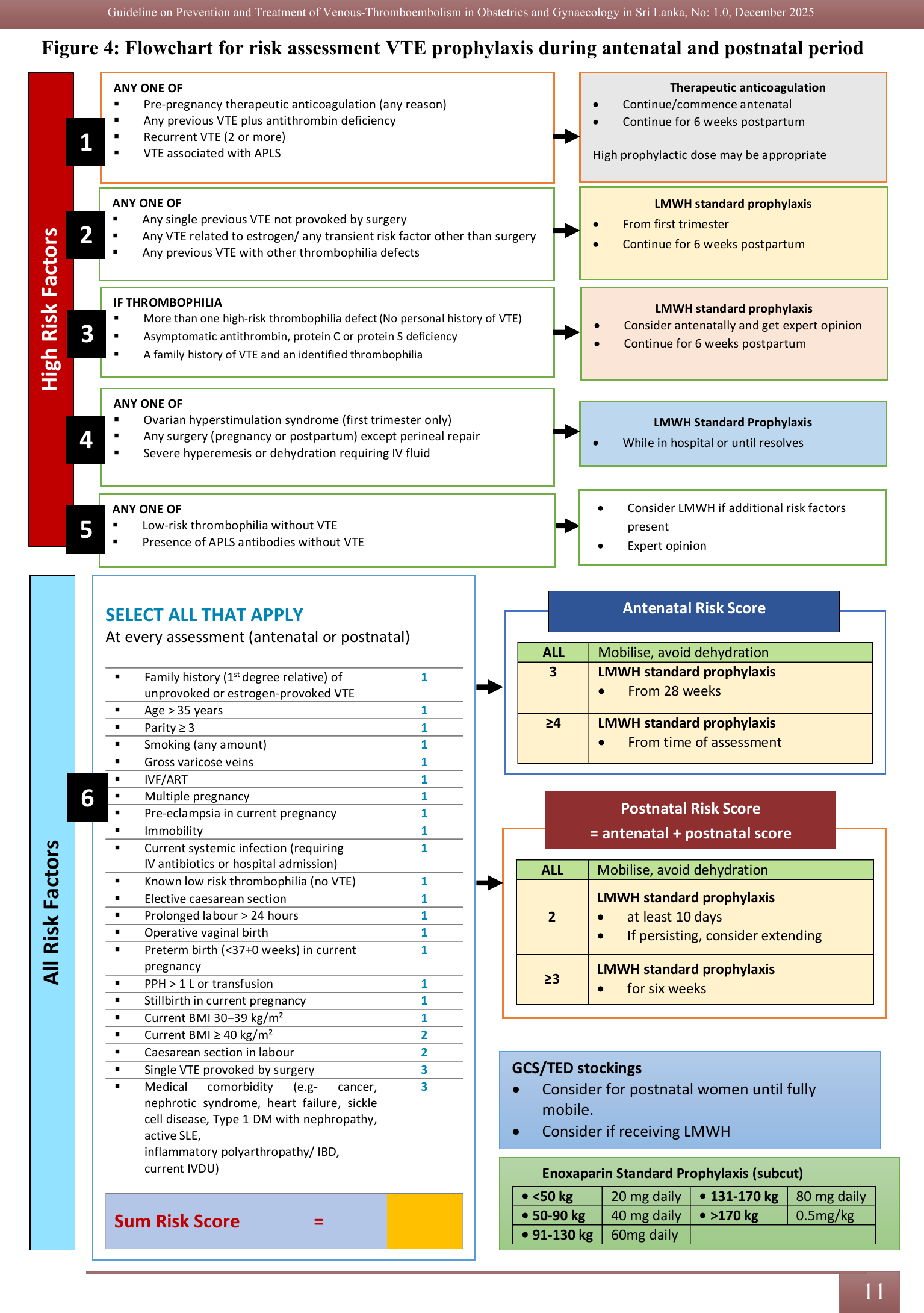
Risk factors can be divided into the following categories as shown in Table 2. All the risk factors can be further stratified according to the degree of risk as shown in Figure 4 (1).
Pre-existing risk factors
Obstetric risk factors: Antenatal & Postnatal
Transient risk factors: These risk factors are potentially modifiable. They may either emerge later in pregnancy or resolve over time.
So, it is essential to conduct continuous, individualised risk assessments throughout the gestational period.
Table 2: Risk factors for VTE in pregnancy
| Pre-existing risk factors | Previous VTE | |
| Thrombophilia | Heritable (2) - Antithrombin deficiency - Protein C deficiency - Protein S deficiency - Factor V Leiden - Prothrombin gene mutation |
|
| Acquired - Antiphospholipid syndrome (3) - Persistent lupus anticoagulant and/or persistent moderate/high titre anticardiolipin antibodies and/or β₂-glycoprotein I antibodies |
||
| Family history of unprovoked or estrogen-related VTE | ||
| Medical comorbidities (e.g., cancer; heart failure; active SLE, inflammatory polyarthropathy or IBD; nephrotic syndrome; type 1 diabetes mellitus with nephropathy; sickle cell disease; current intravenous drug user) | ||
| Age > 35 years | ||
| Obesity (BMI ≥ 30 kg/m²) either pre-pregnancy or at booking | ||
| Parity ≥ 3 | ||
| Smoking | ||
| Gross varicose veins (Symptomatic or above knee or with associated phlebitis, oedema/skin changes) | ||
| Paraplegia | ||
| Obstetric risk factors | Antenatal risk factors:
Multiple pregnancy Assisted reproductive technology (ART) Current pre-eclampsia |
|
| Postnatal risk factors:
Elective or Emergency Caesarean section Prolonged labour (> 24 hours) Mid-cavity or rotational operative delivery Stillbirth Preterm birth Postpartum haemorrhage (> 1 litre/requiring transfusion) |
||
| Transient risk factors | Any surgical procedure in pregnancy or puerperium except immediate repair of the perineum. | |
| Hyperemesis | ||
| Ovarian hyperstimulation syndrome (first trimester only) | ||
| In vitro fertilization (IVF) | ||
| Admission or immobility (e.g. ≥ 3 days bed rest, pelvic girdle pain restricting mobility) | ||
Current systemic infection requiring intravenous antibiotics or admission to hospital (e.g. pneumonia, pyelonephritis) Postpartum wound infection |
||
| Long-distance travel (>4 hours) | ||
Timing of risk assessment
It is recommended to perform risk assessment at following occasions.
Early pregnancy/ At booking visit
At 28 weeks
Repeat assessment if:
Antenatal admission to hospital
Development of pregnancy complication (e.g. pre-eclampsia)
Prolonged immobility
Intrapartum
Postpartum within 6 hours of birth
Care plan on each occasion includes (4):
Discuss options for VTE thromboprophylaxis with woman
Multidisciplinary approach of care: If high risk or any doubtful cases, refer to haematologist for further assessment
Discuss with the anaesthetic team to decide on the peripartum management plan
References
Royal College of Obstetricians and Gynaecologists. Reducing the Risk of Venous Thromboembolism during Pregnancy and the Puerperium: Green-top Guideline No 37A. London, UK: Royal College of Obstetricians and Gynaecologists; 2015 (Ammended 2023)
LockwoodC, WendelG, Silverman N; American College of Obstetricians and Gynaecologists Women's Health Care Physicians. ACOG Practice Bulletin No. 138: inherited thrombophilias in pregnancy. Obstetrics & Gynaecology. 2013;122(03):706-717
Branch DW, Holmgren C, Goldberg JD; Committee on Practice Bulletins-Obstetrics, American College of Obstetricians and Gynaecologists. Practice Bulletin No. 132: antiphospholipid syn drome. Obstetrics & Gynecology.2012;120(06):1514-1521
Deering S, Thiel L, Eubanks A. Risk Assessment and Treatment Guide for Obstetric Thromboprophylaxis: Comprehensive Review of Current Guidelines. American Journal of Perinatology. 2018 Sep 19;36(02):130-5.
4. Guidelines for thrombophilia testing in pregnancy
4.1 General principles in thrombophilia testing
Thrombophilia is an inherited or acquired condition that predispose individuals to thromboembolism. In pregnancy, the hypercoagulable state further increases thrombotic risk (1, 2).
Not all pregnant women require thrombophilia testing.
Testing should be targeted to specific clinical scenarios where results will influence management.
The timing of testing is crucial as pregnancy and anticoagulation can affect test results.
Cost-effectiveness should be considered in the Sri Lankan healthcare setting (3).
Family history of thrombophilia or VTE is an important factor in decision-making.
4.2. Indications for testing
4.2.1 Strong indications (Recommended testing):
Personal history of unprovoked VTE or VTE associated with hormonal therapy
First-degree relative with high-risk thrombophilia (e.g., Antithrombin deficiency, Factor V Leiden homozygosity)
Recurrent pregnancy loss (≥3 first-trimester losses, less than 10 weeks or ≥1 second-trimester loss of a morphological normal fetus)
Previous pregnancy with severe, early-onset pre-eclampsia (<34 weeks) or HELLP syndrome
Previous pregnancy with unexplained FGR (<10th percentile of AC/EFW associated with Doppler abnormalities) or stillbirth
History of arterial thrombosis under age 50 without conventional risk factors
Women fulfilling criteria 3, 4 or 5 with persistently positive APLS antibodies are classified as having Obstetric APLS (4). In this group, anticoagulation is not required for venous thromboembolism prevention. However, Enoxaparin 40 mg daily should be commenced from the time of a positive pregnancy test, continued throughout the pregnancy and up-to 6 weeks postpartum period.
It is also recommended to commence Aspirin 150 mg once daily, from the time of a positive pregnancy test and continue till 36 weeks to improve obstetric outcomes.
4.2.2 Relative indications (Consider testing):
Single late fetal loss (>10 weeks)
Placental abruption with fetal compromise
Family history of an unprovoked or estrogen-provoked VTE in a first-degree relative, aged under 50 years
Severe pre-eclampsia (at any gestational age)
Multiple medical risk factors for VTE during pregnancy
4.2.3 Not recommended:
Routine testing in women with no personal or family history of VTE or adverse pregnancy outcomes (1, 2)
Testing during acute VTE (may give false results)
Universal screening in pregnancy
Testing in women with provoked VTE from major transient risk factors (e.g., major surgery)
4.3. Types of thrombophilia tests
4.3.1 Inherited thrombophilia:
Factor V Leiden mutation
Prothrombin gene (G20210A) mutation
Antithrombin deficiency
Protein C deficiency
Protein S deficiency
4.3.2 Acquired thrombophilia:
Antiphospholipid antibodies:
Lupus anticoagulant (LA)
Anticardiolipin antibodies (aCL) IgG and IgM
Anti-β2-glycoprotein I (aβ2GPI) IgG and IgM (4)
Timing of testing
Ideally performed before pregnancy or at least 3 months after:
Completion of anticoagulation therapy
Resolution of acute thrombotic event
Six weeks are sufficient following delivery or pregnancy loss.
Protein S, protein C, and antithrombin levels are affected by pregnancy, hormonal contraception, and acute thrombosis.
Antiphospholipid antibody testing should be repeated after 12 weeks to confirm persistence if initially positive (4).
Interpretation and management
Positive results should be interpreted within the clinical context
Consider referral to haematologist.
Genetic counselling should be offered for hereditary thrombophilia.
Management decisions should be based on:
Type and severity of thrombophilia
Personal and family history of VTE
Previous pregnancy complications
Additional risk factors for VTE
Practical approach for Sri Lankan setting Testing at resource - limited setting:
Testing at resource - limited setting:
Prioritize testing for women with the strongest indications
Consider a stepwise approach, starting with more common thrombophilias
Reserve comprehensive testing for specialized centres
Referral pathway:
Peripheral health centres should refer to tertiary care for testing
Collaborative management between obstetrics and haematology teams (3)
References
Royal College of Obstetricians and Gynaecologists. Thrombosis and
embolism during pregnancy and the puerperium: reducing the risk.
Green-top Guideline No. 37a. London: RCOG; 2015. American College of Obstetricians and Gynaecologists. ACOG
Practice Bulletin No. 197: Inherited thrombophilias in pregnancy. Obstet
Gynecol. 2018;132(1):e18–34. Sri Lanka College of Obstetricians & Gynaecologists. National
guidelines for management of thromboembolism in pregnancy. Colombo:
SLCOG; 2019. Miyakis S, Lockshin MD, Atsumi T, et al. International consensus
statement on the updated classification criteria for definite
antiphospholipid syndrome (APS). J Thromb Haemost.
2006;4(2):295–306. Confirm DVT by imaging (Refer chapter 6) D-Dimer physiologically increases during the pregnancy, but has a
high negative predictive value (1) Following baseline investigations should be carried out prior to
commence treatments (2): PT/INR, APTT FBC Serum Creatinine LFT (AST/ALT) Booking body weight should be considered for dose calculation
(3) Special treatment may be required based on individual basis such
as, Embolectomy Thrombectomy Thrombolytic therapy Follow Table 3 for different treatment options at
various clinical scenarios. Commence Heparin. Available options are Low Molecular Weight Heparin (LMWH) and High
Molecular Weight Heparin (HMWH)/ Unfractionated Heparin. Confirm diagnosis by investigations (Compression duplex
ultrasound). Continue the treatments as given below; Suspicious Doppler, but not confirmed DVT Stop Heparin Repeat scan in 3 days & 7days if high level of clinical
suspicion exists (4). However, if there is a persistent clinical suspicion, treatment
should be continued and repeat scan in 3 days & 7days later. If low level of suspicion treatments should be stopped. LMWH 1mg/ kg BD (Body weight at booking visit) OR 1.5 mg/kg once
daily. Doses should be round-off to vial size. HMWH: 5000 IU bolus followed by 18IU/kg/hr infusion Monitor APTT, maintain 1.5 - 2 times normal Check Platelet count between 4 -14 days once in 3 days Lower doses of LMWH (Enoxaparin and Dalteparin) should be
employed if the creatinine clearance is less than 30 ml/minute or
Tinzaparin if less than 20 ml/minute. Renal dose of LMWH is usually 20-40 mg daily (Obtain expert
opinion). HMWH – Doses are as above. Intravenous unfractionated heparin Expert advice and thrombolysis - Thrombolytic therapy or thoracotomy
and surgical embolectomy Special treatment options (Refer chapter 6) Thoracotomy, embolectomy and thrombolytic therapy on individual
basis. IVC Filter – indicated when there are recurrent PE and thrombus
involving Iliac veins. DVT at term: Continue HMWH infusion till 6 hours prior to expected time of
delivery (5) LMWH treatment dose should be stopped 24 hrs prior to
delivery LMWH should not be given for 4 hours after the use of spinal
anaesthesia or after the epidural catheter has been removed (6,7). Epidural catheter should not be removed within 12 hours of the most
recent injection. In the initial management of DVT, the leg should be elevated and
a graduated elastic compression stocking applied to reduce oedema.
(controversial evidence) Mobilisation with graduated elastic compression stockings should
be encouraged When there is a high risk of haemorrhage Post partum period to be managed with Avoid warfarin till 5th day of post-partum period or
longer in case of bleeding Post Thrombotic Syndrome to be avoided by Wearing of stockings (controversial evidence) Use of anticoagulant for 3 months Thrombophilia screening should be planned with haematologist
(Refer chapter 4). 1. American Society of Haematology. ASH guidelines for VTE
prophylaxis and treatment in pregnancy. Washington (DC): ASH; 2018.
Available from: (https://gynaecolo.hematology.org) 2. Royal College of Obstetricians and Gynaecologists. Reducing the
risk of venous thromboembolism during pregnancy and the puerperium
(Green-top Guideline No. 37a). London: RCOG; 2015. 3. Royal College of Obstetricians and Gynaecologists. Green-top
Guideline No. 37a: LMWH dosing based on booking weight and anticoagulant
dosing guidance. London: RCOG; 2015. 4. Ministry of Health / institutional consensus group. Doppler-based
protocol for suspected deep venous thrombosis in pregnancy: repeat
imaging at 3 and 7 days. Kampala: Uganda Ministry of Health; Year not
stated. 5. UpToDate. Neuraxial anaesthesia in anticoagulated pregnant
patients. Waltham (MA): UpToDate Inc.; [cited 2025]. Available from:
[https://gynaecolo.uptodate.com] (https://gynaecolo.uptodate.com) 6. Horlocker TT, Vandermeulen E, Kopp SL, et al. Regional anaesthesia
in the anticoagulated patient: ASRA and Scandinavian guidelines. Reg
Anesth Pain Med. Year varies by edition. 7. Stanford University School of Medicine. Neuraxial anticoagulation
guideline: LMWH and neuraxial anesthesia timing recommendations.
Stanford (CA): Stanford Medicine; Year not stated. 8. Greer IA, Nelson-Piercy C. Venous thromboembolism in pregnancy. Am
Fam Physician. 2008;77(12):1709–16. Early radiological diagnosis and interventional procedures play a critical role in preventing fatal complications such as PE in these high-risk categories (1, 2). In Sri Lanka, there is a growing need for a standardised approach to early radiological detection and intervention to minimise mortality and improve patient outcomes in obstetrics and gynaecology (3, 4). The primary imaging modality for diagnosing DVT is Compression Ultrasound Scan (CUS) with Doppler (1, 3). Pelvic or iliac vein thrombosis is often missed on standard ultrasound, especially after gynaecological cancer surgery or in pregnancy. In pregnancy, Magnetic Resonance Venography (MRV) is recommended. (1, 4). For non-pregnant gynaecological patients, CT venography is recommended to diagnose post-surgical pelvic DVT, particularly in cases of ovarian or endometrial cancer-related thrombosis (2). While Contrast Venography remains the gold standard, it is rarely used due to its invasive nature (5). For suspected PE, CT Pulmonary Angiography (CTPA) is the gold standard due to its high sensitivity and specificity, especially when CXR is abnormal (1,5). CTPA enables rapid and accurate detection of pulmonary thromboembolism. It can also identify alternative diagnoses such as pneumonia or amniotic fluid embolism (4). While radiation exposure is a concern in pregnancy, modern low-dose protocols and proper shielding techniques significantly minimize fetal radiation risks (3). Ventilation–Perfusion (V/Q) scanning remains the preferred first-line imaging modality for suspected pulmonary embolism (PE) in pregnancy when the chest X-ray is normal. V/Q scanning provides lower maternal breast radiation exposure. However, the availability of V/Q scanning across Sri Lankan tertiary and secondary care centres is extremely limited, resulting in constrained real-world applicability of this recommendation. This guideline therefore adopts a context-aligned, safety-driven escalation pathway: For high-risk patients who cannot receive anticoagulation, interventional radiology provides life-saving treatment options (1, 2). These include: Emerging radiological and intervention techniques offer safer and more effective options for diagnosing and managing thrombosis, including (2, 4): Due to the limitations of some facilities in Sri Lanka, direct engagement with the interventional radiologist/ radiologist early is recommended to support clinical decision-making. Pregnant women with Mechanical Heart Valves (MHVs) face significant challenges due to their high thrombotic risk combined with the hypercoagulable state of pregnancy (1-3). Management requires balancing maternal thromboembolism risk against risks of bleeding and adverse fetal outcomes (3,4). Although mechanical-valve thrombosis differs pathophysiologically from VTE, the guideline committee recognised its unique pregnancy anticoagulation requirements and therefore included it as a separate dedicated section. Option 1: Vitamin K Antagonists (VKA) throughout pregnancy (5,6) Option 2: Low Molecular Weight Heparin (LMWH) throughout pregnancy Option 3: Sequential therapy Planned vaginal delivery Caesarean section Valve thrombosis (10) Haemorrhage 7.2. Management of superficial vein thrombophlebitis (SVT) in pregnancy In pregnancy, superficial vein thrombophlebitis (SVT) is characterised by the formation of a thrombus and inflammation in a superficial vein, typically in the lower extremities (1). Although SVT is typically self-limiting, it can progress to the deep venous system, which elevates the likelihood of pulmonary embolism (PE) and deep vein thrombosis (DVT) (2). Symptoms are frequently unilateral and typically manifest in the great saphenous vein or small saphenous vein. Localised tenderness, redness, warmth, and oedema in a superficial vein can be elicited in examination (4). Thickening of the afflicted vein become palpable and resembles a cord. There is no substantial leg swelling, which is more indicative of DVT (2). Warning signs of DVT or PE include: • Sudden swelling and pain throughout the entire limb • Tachycardia, chest pain, and shortness of breath • Haemodynamic instability or cyanosis (5). The diagnosis is predominantly clinical, based on localised inflammation along the superficial vein (1). Compression Doppler ultrasound (USG): • Necessary when there is a suspicion of extension towards deep vessels. • Aids in the differentiation of SVT from DVT, particularly in cases of symptomatic DVT (4). D-dimer test: • The levels are physiologically elevated, rendering them unreliable during pregnancy. • Utilised exclusively when systemic coagulopathy or thrombophilia is suspected (6). 7.2.4. Management 7.2.4.1 Conservative management (Uncomplicated SVT, localised <5 cm) • Compression stocking (Class II or III, 20–30 mmHg) to enhance venous return (2). • Elevate the legs to alleviate discomfort and oedema. • Promote ambulation (avoid prolonged periods of bed rest). • Compresses that are either warm or chilly are employed to alleviate discomfort. • Paracetamol or NSAIDs (e.g., Ibuprofen) for pain relief (NSAIDs should be avoided in the third trimester) (5). 7.2.4.2 Anticoagulation therapy (For high-risk cases or extensive SVT) Anticoagulation indicates if: • SVT with a length of at least 5 cm Anticoagulants that are advised during pregnancy include: • Low molecular weight heparin (Preferred choice): It is not recommended to use Direct Oral Anticoagulants (DOACs) during pregnancy, such as Apixaban and Rivaroxaban (5). The prevalence of VTE after gynaecologic surgery varies from 15% to 30%. Fatal PE occurs in 0.2–0.9% of patients. Risk factors for VTE in gynaecological patients are depicted in Table 04. The risk of VTE after gynaecologic laparoscopic surgery is uncertain (1,2). The American College of Chest Physicians (ACCP) originally incorporated the Caprini Risk Assessment Model, where each independent risk factor is weighted from 1 to 5 points, and the cumulative score drives the intensity of thromboprophylaxis required (1). A 2022 update published in The Obstetrician & Gynaecologist (TOG) re-evaluated the Caprini model against NICE, RCOG, ACOG, and ACCP guidance and produced a modified version tailored specifically for gynaecological patients (3). For national standardisation, this guideline adopts the validated TOG-modified chart to support operational decision-making in routine gynaecological practice (Table 4). It is recommended by the guideline committee to use Table 4 and Table 5 for every patient undergoing gynaecological surgery and attach to the Bed Head Tickets. Table 4: Risk factors and scoring Table 5: Summary on recommended thromboprophylaxis methods for various risk stratifications in gynaecology Abbreviations for the table 5: VTE = venous thromboembolism; IPC = intermittent pneumatic compression; GCS = graduated compression stockings; LMWH = low-molecular-weight heparin. Refer Table 5 for recommended thromboprophylaxis methods for various risk scores (3). Table 6 highlights the various pharmacological options for thromboprophylaxis in gynaecology (4-6). Table 6: Summary of different pharmacological prophylaxis agents used in gynaecological thromboprophylaxis Patients with cancer have up to a 7-fold increased risk of venous thromboembolism (VTE) compared to the general population (1, 2). Among gynaecological oncology patients, the risk is even more significant, with approximately 38% experiencing perioperative thromboembolic complications in the absence of prophylaxis (3). This elevated risk is attributed to multiple cancer-related factors, including the malignancy itself, advanced age, vascular compression by pelvic masses, prolonged surgical procedures, vascular injury and chemotherapy-induced hypercoagulability (4). Ovarian cancer has one of the highest VTE incidence rates among gynaecological malignancies, with reported rates ranging from 5% to 16.6% (5). Risk factors for early VTE include surgery and chemotherapy. Later VTE events are associated with older age, prior DVT, advanced FIGO stage (IIC–IV), and residual tumour following primary surgery (6). This is one of the most prevalent gynaecologic cancers. Prominent VTE risk factors include metabolic syndrome components such as obesity, hypertension, insulin resistance, diabetes, and dyslipidaemia (7). Treatment modalities in oncology further contribute to thrombosis risk. These include: Each of these factors has been independently associated with an increased likelihood of developing VTE. All women undergoing gynaecological surgery including those with gynaecological malignancies should be risk-stratified using the TOG-modified Caprini Risk Assessment Model (2022), which has been benchmarked against NICE, RCOG, ACOG, and ACCP frameworks and validated for gynaecological practice. This model is adopted as the standardised national tool for operational VTE risk assessment in Sri Lanka (Table 4). It is recommended by the guideline committee to use Table 4 and Table 5 for every patient undergoing surgery for gynaecological malignancies and attach to the Bed Head Tickets. In addition to that, The American College of Chest Physicians (ACCP) recommends extended-duration pharmacologic prophylaxis (i.e., 4 weeks of LMWH) for women undergoing gynaecological cancer surgery who are at high risk of VTE and not at elevated risk for bleeding (10). Individualised risk assessment and expert opinion should be taken for extended duration of pharmacological prophylaxis. Despite limited data regarding minimally invasive surgery (MIS), it is recommended to follow the same thromboprophylaxis guidelines as for laparotomy. Patients receiving chemotherapy or antiangiogenic agents face a heightened risk of VTE. In such cases, Low Molecular Weight Heparin (LMWH) is the preferred anticoagulant due to its efficacy and safety profile in cancer patients. 1. Zawilska K, Bała MM, Błędowski P, Chmielewski DW, Dobrowolski Z, Frączek M, et al. [Polish guidelines for the prevention and treatment of venous thromboembolism. 2012 update]. Pol Arch Med Wewn. 2012;122 Suppl 2:3–74. 2. Khorana AA, Francis CW, Culakova E, Kuderer NM, Lyman GH. Frequency, risk factors, and trends for venous thromboembolism among hospitalized cancer patients. Cancer. 2007 Nov 15;110(10):2339–46. 3. Lyman GH, Khorana AA, Kuderer NM, Lee AY, Arcelus JI, Balaban EP, et al. Venous Thromboembolism Prophylaxis and Treatment in Patients With Cancer: American Society of Clinical Oncology Clinical Practice Guideline Update. J Clin Oncol. 2013 Jun 10;31(17):2189–204. 4. Barber EL, Clarke-Pearson DL. The limited utility of currently available venous thromboembolism risk assessment tools in gynecological oncology patients. Am J Obstet Gynecol. 2016 Oct;215(4):445.e1-445.e9. 5. Schmeler KM, Wilson GL, Cain K, Munsell MF, Ramirez PT, Soliman PT, et al. Venous thromboembolism (VTE) rates following the implementation of extended duration prophylaxis for patients undergoing surgery for gynecologic malignancies. Gynecol Oncol. 2013 Feb;128(2):204–8. 6. Duska LR, Garrett L, Henretta M, Ferriss JS, Lee L, Horowitz N. When ‘never-events’ occur despite adherence to clinical guidelines: The case of venous thromboembolism in clear cell cancer of the ovary compared with other epithelial histologic subtypes. Gynecol Oncol. 2010 Mar;116(3):374–7. 7. Matsuo K, Yessaian AA, Lin YG, Pham HQ, Muderspach LI, Liebman HA, et al. Predictive model of venous thromboembolism in endometrial cancer. Gynecol Oncol. 2013 Mar;128(3):544–51. 8. Sedlis A, Bundy BN, Rotman MZ, Lentz SS, Muderspach LI, Zaino RJ. A Randomized Trial of Pelvic Radiation Therapy versus No Further Therapy in Selected Patients with Stage IB Carcinoma of the Cervix after Radical Hysterectomy and Pelvic Lymphadenectomy: A Gynecologic Oncology Group Study. Gynecol Oncol. 1999 May;73(2):177–83. 9. Bouchard-Fortier G, Geerts WH, Covens A, Vicus D, Kupets R, Gien LT. Is venous thromboprophylaxis necessary in patients undergoing minimally invasive surgery for a gynecologic malignancy? Gynecol Oncol. 2014 Aug;134(2):228–32. Incidence can rise up to 1.7% if developed ovarian hyperstimulation syndrome (OHSS) with hospitalization (2) Refer the Table 7 for recommendations adopted from the Swedish Association of Obstetrics and Gynaecology (SFOG) . It is recommended by the guideline committee to use this chart (Table 7) to risk stratify every patient undergoing IVF and attach this to the Bed Head Tickets. Table 7: Recommendations adopted from the Swedish Association of Obstetrics and Gynaecology (SFOG) for thromboprophylaxis in IVF In addition to that, it is recommended to follow the guiding principles given below. 1.Rova K, Passmark GYNAECOLO, Lindqvist PG. Venous thromboembolism in relation to in-vitro fertilization: an approach to determining the incidence and increase in risk in successful cycles. Fertil Steril. 2012;97(1):95-100. 2.Nelson SM. Venous thrombosis during assisted reproduction: novel risk-reduction strategies. Thromb Res. 2013;131(Suppl 1):S1-S3. doi:10.1016/S0049-3848(13)00023-6. 3. Swedish Society of Obstetrics and Gynecology (SFOG). Guideline for thromboprophylaxis during in vitro fertilisation (IVF). Stockholm: SFOG; 2018.
5. Acute management of thromboembolism in pregnancy
5.1. General facts
5.2. Treatments
Table 3: Treatment options at various clinical scenarios
Clinical Scenario
Treatment Options
Suspect DVT
Treatment
(When baseline investigations are normal)
Treatment
(When serum creatinine is high)
Massive PE
Treatment maintenance
Therapeutic dose of LMWH should continue throughout pregnancy and 6
weeks post-partum or up to at least 3 months in total (1, 2).
Deciding on Heparin closer to delivery
When patient is with epidural OR had a spinal anaesthesia
Special facts
Heparin infusion is preferred.
Warfarin or LMWH, as they are not contraindicated during breast feeding
(1,8)
References
6. Role of radiology in diagnosis, treatment and prevention
6.1. Introduction
6.2. Early radiological diagnosis of Deep Vein Thrombosis (DVT)
6.3. Radiological diagnosis of Pulmonary Embolism (PE)
6.4. Interventional radiological options to prevent PE in high-risk patients
6.5. Newer advances in radiological thromboprophylaxis
References
7. Special circumstances in obstetrics
7.1. Thromboprophylaxis for pregnant women with mechanical heart valves
7.1.1. Pre-conception counselling
7.1.2. Anticoagulation regimens during pregnancy
Disadvantages:
Disadvantages:
LMWH during 6-12 weeks of gestation
7.1.3. Specific recommendations for each regimen
7.1.3.1 Vitamin K antagonist (Warfarin) regimen (7,8)
7.1.3.2 LMWH regimen
7.1.3.3 Unfractionated Heparin (UFH) regimen
7.1.4. Risk stratification and personalized approach
High-risk patients (ANY of the following):
LVEF <35%
Risk-adapted recommendations:
7.1.5. Delivery planning
7.1.6. Postpartum management
VKA started 5 days post-delivery if no bleeding complications.
7.1.7. Additional considerations for Sri Lanka
7.1.8. Management of complications
References
7.2.1. Risk factors for superficial vein thrombophlebitis in pregnancy Following are risk factors for SVT during pregnancy:
7.2.2. Clinical presentation
7.2.3. Diagnosis
Blood tests:
References
8. Thromboprophylaxis in gynaecological patients
8.1. Introduction
Risk Factor Score Risk Factor (Patient-Related) Risk Factor (Patient-Related) Age 41–60 years +1 61–74 years +2 ≥75 years +3 BMI ≥30 +1 ≥40 +2 COCP/oral HRT use +1 Pregnancy or within 6 weeks following delivery/miscarriage +3 Varicose veins +1 Pelvic mass of a significant size (e.g., fibroid >12 weeks in size) +1 Personal VTE history +3 Family history of unprovoked VTE +3 Known high-risk inherited thrombophilia +3 Currently on bed rest (inability to walk 10 meters) +1 Patient confined to bed >72 hours +2 Medical conditions (acute MI, COPD, Inflammatory bowel disease, Heart failure, Sepsis, pneumonia, diabetes on insulin) +1 Major surgery in the past month +1 Hip, pelvis or leg fracture; stroke; multiple trauma; acute spinal cord injury +5 Past/present cancer +2 Smoking +1 Immobilizing plaster cast +2 Blood transfusion +1 Risk Factor (Procedure-Related) Risk Factor (Procedure-Related) Duration of surgery (From anaesthesia to end of surgery) <60 minutes +1 Duration of surgery >60 minutes +2 Total Score Risk score Risk stratum Thromboprophylaxis 0 Low risk Ambulation alone or IPC or GCS during hospitalisation 1–4 Moderate risk IPC ± GCS during hospitalisation 5–8 High risk IPC + LMWH or LMWH alone for 7–10 days ≥9 Highest risk IPC + LMWH or LMWH alone for 30 days 8.2. Methods of thromboprophylaxis in gynaecology
8.2.1. Mechanical prophylactic measures
8.2.2. Pharmacological prophylaxis
Agent Dose Advantages Disadvantages / Considerations Low Molecular Weight Heparin (LMWH) Enoxaparin: 40 mg SC (0.5mg/kg) once daily daily
Dalteparin: 5000 IU SC once daily Once-daily dosing
Greater bioavailability
Lower risk of HIT compared to UFH Requires renal dose adjustment
Local injection site reactions
Rare risk of HIT Unfractionated Heparin (UFH) 5000 IU SC every 8–12 hours Effective
Shorter half-life
Easier perioperative control Requires frequent dosing
Higher risk of bleeding
HIT (up to 6% incidence) References
9. Special circumstances in gynaecology
9.1. Thromboprophylaxis in gynaecological malignancies
9.1.1. Increased VTE risk in gynaecological malignancies
9.1.2. Tumour-specific VTE risk
Ovarian cancer:
Endometrial carcinoma:
Cervical Cancer:
VTE risk in cervical cancer varies widely from 0% to 34%, with the highest rates observed in mucin-producing tumours (8).
9.1.3. Impact of oncological treatments on VTE risk
9.1.4. Recommendations for prophylaxis
9.1.5. VTE risk with chemotherapy and antiangiogenic therapy
References
10. Clarke-Pearson DL, Abaid LN. Prevention of Venous Thromboembolic Events After Gynecologic Surgery: Obstet Gynecol. 2012 Jan;119(1):155–67.
9.2. Thromboprophylaxis in assisted reproduction
VTE occurrence is estimated at approximately 1 per 1,000 in spontaneous pregnancies, rising to around 2 per 1,000 in pregnancies following IVF (1).
OHSS affects about 5–7% of IVF cycles, with severe forms accounting for up to a 100-fold increased risk of VTE (3).
9.2.2. Recommendations for thromboprophylaxis in IVF
Score Risk Factors 1 Heterozygous FV Leiden
Heterozygous prothrombin mutation
Obesity (BMI >28 at booking)
Caesarean section
Hereditary factors: VTE in a 1st degree relative < 60 years
Age >40 years
Preeclampsia
Hyperhomocysteinemia
Placental abruption
Inflammatory bowel disease 2 Protein S deficiency
Protein C deficiency
Immobilization (strict immobilization or if the patient has a cast) 3 Homozygous FV Leiden
Homozygous prothrombin
More than one thrombophilia defect 4 Prior VTE
APLS without VTE
OHSS Very high Risk Mechanical aortic valve
Condition warranting continuous thromboprophylaxis
APLS with VTE
Recurrent VTE
Antithrombin deficiency Treatment according to score: Treatment according to score: 1 point Thromboprophylaxis not needed 2 points Thromboprophylaxis postpartum once daily for at least 7 days, this includes thromboprophylaxis for a transient risk factor 3 points Thromboprophylaxis once daily for 6 weeks postpartum 4 or higher points Thromboprophylaxis once daily throughout pregnancy and at least for 6 weeks postpartum Very high risk Thromboprophylaxis twice daily (double dose) throughout pregnancy and at least for 12 weeks postpartum References
Patient category Quick reference Thromboprophylaxis in all antenatal and postnatal women Figure 4 Acute management of thromboembolism in pregnancy in various clinical scenarios Table 3 All gynaecological patients Table 4 and Table 5 Thromboprophylaxis in gynaecological malignancies Table 4 and Table 5
Expert opinion for extended use Thromboprophylaxis in assisted reproduction Table 7