
Background
The evaluation of fetal growth is one of the critical objectives in perinatal care. Impaired fetal growth is a leading cause of stillbirth, neonatal mortality, and neonatal morbidity, both short- and long-term, and increases the risk of non-communicable diseases in adulthood.
This guideline provides the available evidence and recommendations regarding prenatal prediction, prevention, diagnosis, monitoring and management options for pregnancies complicated with SGA fetuses/ FGR, with the overall goal of decreasing fetal and neonatal morbidity and mortality.
Definitions
SGA is defined as birthweight below the 10th centile for gestational age. Antenatally SGA fetus can be identified by either EFW or AC <10th centile for gestational age. The majority of SGA fetuses (70%) are constitutionally small, and the remaining (30%) are growth restricted, where the fetus fails to reach its genetically predetermined growth potential.
Placental-mediated causes account for 70% of FGR, and the rest are due to non-placenta-mediated causes such as structural or chromosomal anomaly, inborn errors of metabolism and fetal infection. As an EFW of less than the 3rd centile is associated with the highest risk for stillbirth and perinatal mortality, when diagnosing FGR, AC or EFW below the 3rd centile can be used as an isolated criterion to define FGR at any gestational age.
A consensus-based definition to classify placenta-mediated FGR has been proposed via a Delphi procedure. It is based on a combination of fetal size measurement (AC and EFW) and abnormal Doppler findings in the umbilical, uterine and middle cerebral arteries, as described in Table 1.
Early-versus late-onset FGR
Placenta-mediated FGR is classified into early (<32 weeks) and late (32 weeks) FGR based on the gestational age at diagnosis1.
Early onset FGR has a prevalence of 30%, is usually more severe, and is more likely to be associated with abnormalities in the umbilical artery, middle cerebral artery, and ductus venosus Doppler. The underlying placental pathology includes poor placental implantation, spiral artery abnormalities and associated maternal vascular mal-perfusion similar to early onset pre-eclampsia, leading to its strong association with pre-eclampsia. Although it is easier to detect, the main challenge lies in the delivery timing, balancing the risks of stillbirth and prematurity.
Late-onset FGR is more common, accounting for 70% of placenta-mediated FGR. It is usually milder and associated with normal umbilical artery Doppler with cerebro-placental redistribution. The underlying pathophysiology includes altered placental diffusion, therefore, is less likely to be associated with pre-eclampsia. The main challenge lies in the diagnosis. Given that the Doppler studies are normal in late-onset FGR, the natural history is less predictable, with a risk of sudden decompensation and stillbirth if not diagnosed early.
Aetiology of SGA/FGR
Common aetiologies of FGR are listed in Table 2.
Table 2. Etiologies of FGR
| Suboptimal uteroplacental perfusion of fetal nutrition |
|---|
| a. Pre-placental (maternal) factors |
| Hypoxaemia (chronic lung disease, high altitude) Anaemia Smoking, substance abuse (cocaine, methamphetamines) Malabsorption, poor weight gain Environmental toxins: air pollution, heavy metals (lead, mercury), perfluorooctanoic acid (PFOA) |
| b. Placental factors |
| Maternal vascular mal-perfusion pathology (infarction, fibrin deposition, chronic abruption) Fetal vascular mal-perfusion pathology Chronic placental inflammation (villitis of unknown aetiology) Confined placental mosaicism |
| c. Post-placental (umbilical cord) factors |
| Increased coiling Increased cord length True cord knot Single umbilical artery Marginal or velamentous cord insertion |
| Fetal disorders |
| Genetic: chromosomal, microdeletions/ duplications, single-site mutations, epigenetic disorders Structural abnormalities: congenital heart disease, gastroschisis Congenital infections: cytomegalovirus, toxoplasmosis, herpes, rubella, syphilis, Zika virus, malaria, HIV Teratogen exposure: drugs, toxins |
Risks associated with SGA/FGR
FGR or SGA account for up to 30% of antepartum stillbirths. It contributes significantly to iatrogenic preterm delivery and is an independent risk factor for spontaneous preterm birth. Despite improvements in neonatal care, FGR is significantly associated with neonatal mortality and both short- and long-term neonatal morbidity. Perinatal mortality is five to ten-fold higher in term FGR fetuses compared to appropriately grown neonates.
Among preterm infants, the co-existence of FGR further worsens the risks of prematurity-related compli-cations such as respiratory distress, intraventricular haemorrhage, necrotizing enterocolitis, and metabolic derangements. Among term infants, FGR increases the risk of low cord arterial pH, low Apgar score, and neonatal complications such as hypoglycaemia, hypothermia, and jaundice.
FGR infants are also at increased risk of adverse long-term neurodevelopment outcomes. Low birth weight has been associated with an increased risk of future non-communicable diseases, including obesity, diabetes, hypertension, and cardiovascular disease (Barker hypothesis).
Table 3 summarizes the risks associated with FGR.
Table 3. Risks associated with fetal growth restriction
| Antenatal |
|---|
| Stillbirth Pre-eclampsia Placental abruption Preterm birth |
| Neonatal (short-term) |
| Neonatal mortality Neonatal morbidity: hypoglycemia, hyperbilirubinemia, hypothermia, necrotising enterocolitis, respiratory morbidity, intraventricular haemorrhage |
| Neonatal (long-term) |
| Neurodevelopmental disorders Metabolic syndrome: obesity, hypertension, diabetes, cardiovascular disease. |
Early prediction of SGA/FGR
Early prediction can identify women at high risk of FGR who may benefit from preventive interventions and close monitoring during pregnancy. Methods for early prediction of SGA include history-based screening, screening with serum biochemical markers, and ultrasound markers1.
History-based screening
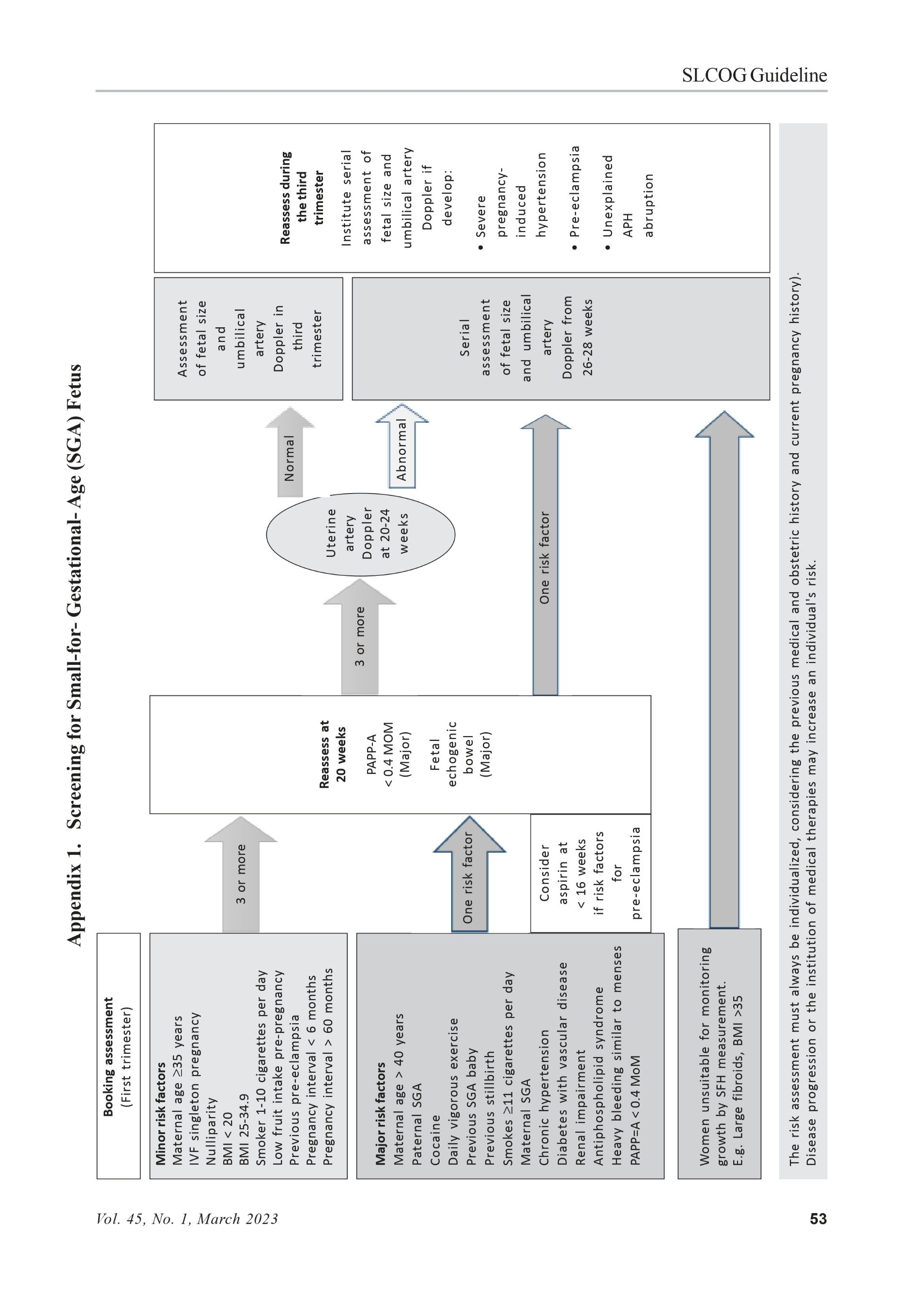
All women should be assessed at booking for risk factors for FGR (Table 4) to identify those who need increased surveillance. Women with a major risk factor should be offered serial ultrasound measurement of fetal size and assessment of fetal wellbeing with umbilical artery Doppler from 26-28 weeks of gestation. Women with three or more minor risk factors should be offered uterine artery Doppler at 20-24 weeks of gestation. Those with normal uterine artery Doppler should be offered an assessment of the fetal size and umbilical artery Doppler in the third trimester, while those with abnormal uterine artery Doppler should be offered serial assessment of fetal size and umbilical artery Doppler from 26-28 weeks’ gestation (Appendix 1).
Table 4. Risk factors for SGA
| Major risk factors: | Minor risk factors: |
|---|---|
| Maternal age > 40 years Smokes 11 cigarettes per day Paternal SGA Cocaine Daily vigorous exercise Previous SGA baby Previous stillbirth Maternal SGA Chronic hypertension Diabetes with vascular disease Renal impairment Antiphospholipid syndrome Heavy bleeding similar to menses PAPP-A < 0.4 MoM Fetal echogenic bowel (at 20 weeks) | Maternal age 35 years IVF singleton pregnancy Nulliparity BMI < 20 BMI 25-34.9 Smokes 1-10 cigarettes per day Low fruit intake pre-pregnancy Previous pre-eclampsia Pregnancy interval <6 months Pregnancy interval 60 months |
Biochemical markers
There is no role for routine screening with serum biochemical markers for FGR. However, when biochemical markers are used as a part of screening for trisomy 21, they may be used for risk stratification for FGR.
A low level of PAPP-A (<0.415 MoM) is considered a major risk factor for an SGA neonate and indicates close surveillance of fetal growth.
Elevated maternal serum levels of alpha-fetoprotein (AFP) in the 2nd trimester, elevated human chorionic gonadotropin (hCG) levels greater than 2.5 MoM in the 2nd trimester, and placental growth factor (PlGF) have limited predictive accuracy for their routine use in clinical practice.
Ultrasound markers
- Uterine artery Doppler
First-trimester uterine artery Doppler has a low sensitivity (12-25%) for SGA neonates, although it has high specificity (91-96%) and high negative predictive value (91-99%). Therefore, it cannot be recommended for routine screening in low or high-risk pregnancies2.
Uterine artery Doppler at 20-24 weeks of gestation has a moderate predictive value for a severely FGR neonate in high-risk populations. For women with three or more minor risk factors, uterine artery Doppler should be offered at 20-24 weeks where resources and expertise are available. Those with abnormal uterine artery Doppler at 20-24 weeks (PI >95th centile and/or notching) should be offered serial ultrasound measurements of fetal growth and wellbeing with umbilical artery Doppler from 26-28 weeks’ gestation2.
Fetal echogenic bowel
As fetal echogenic bowel is independently associated with SGA and fetal demise, serial ultrasound measurement of growth and umbilical Doppler is recommended following a diagnosis of fetal echogenic bowel.
Prevention of SGA/FGR
Lifestyle modification
Ideally, all women should plan their pregnancies, be advised on healthy lifestyles, and optimize their medical conditions and body mass index.
Monitoring of body mass index and advising on target weight gain should be individualized, as insufficient gestational weight gain is associated with an increased risk of FGR.
Substance use, including smoking, alcohol, and illicit drugs, is associated with low birth weight and increased perinatal morbidity and mortality risk. Smoking cessation at any point in pregnancy is beneficial, with the most significant benefit being for cessation before 15 weeks. The risk of SGA with alcohol is increased with as little as one drink per day. Alcohol and illicit drug cessation should be advised.
Medical interventions
- Aspirin
Aspirin is recommended for women at increased risk of pre-eclampsia, with evidence showing that it may reduce the risk of FGR by nearly half if started from 12 weeks onwards until 36 weeks, preferably before 16 weeks of gestation. Higher dosages of aspirin are associated with a greater reduction, favouring a dose of 100-150 mg. Administration of Aspirin in the evening is associated with greater reduction of FGR and pre-eclampsia3.
Progesterone and calcium
There is no evidence that either progesterone or calcium is effective in reducing the incidence of FGR/SGA neonates4,5.
- Low molecular weight heparin (LMWH) Based on the most up-to-date evidence, LMWH cannot be recommended to prevent FGR in women at high risk of placenta-mediated complications6.
Diagnosis of SGA/FGR
Abdominal palpation:
Abdominal palpation has limited accuracy for detecting SGA neonate (sensitivity 19-21%, specificity 98%) and, therefore, should not be routinely performed in the detection of SGA/FGR.
Symphysis-fundal height (SFH) measurement:
SFH is defined as the distance from the top of the uterine fundus (variable point) to the upper border of the pubis symphysis (fixed point) in centimetres. It is measured in the supine position using non-elastic metric tape after the pregnant woman empties her bladder.
Measurement of SFH is the primary screening tool for FGR in low-risk pregnancies. However, it has limited accuracy in predicting SGA, with a sensitivity of 58% and specificity of 87%. There is significant intra- and inter-observer variation with SFH measurements, with serial measurement improving its predictive accuracy. Women with a single SFH which plots below the 10th centile or serial measurements which demonstrate slow or static growth (i.e., they cross centiles in a downward direction) should be referred for ultrasound evaluation. Maternal obesity, abnormal fetal lie, large fibroids, polyhydramnios and engaged fetal head, contribute to the limited predictive accuracy of SFH.
Customized charts (adjusted for maternal characteristics such as maternal weight, height, parity and ethnic group) may improve the detection of SGA neonates; however, no customized SFH charts exist for the Sri Lankan population7.
Sonographic fetal weight estimation
Sonographic fetal biometry includes the assessment of biparietal diameter (BPD), head circumference (HC), abdominal circumference (AC), and femur length (FL). Fetal weight is estimated based on these indices using various equations. The accuracy of most equations falls within ±10%, and the error is greater at the extremes of fetal weight and also affected by fetal sex, presentation, and multiple pregnancies. Equations based on 3-4 biometric indices provide the most accurate weight, with the equation varying among different populations. While it is reasonable to choose an equation validated within the local population, in the absence such a validated equation for the Sri Lankan population, we recommend usage of the Hadlock 3/4, (BPD, HC, AC, and FL) as it provides the greatest accuracy8.
Role of routine third-trimester ultrasound to assess fetal growth
The pregnancy outcome prediction (POP) study comparing the detection of SGA by routine ultrasound versus clinically indicated ultrasound in the 3rd trimester showed a three-fold higher detection rate of SGA in the routine third-trimester ultrasound group (57% vs 20%). Therefore, it is recommended to perform a routine 3rd trimester ultrasound scan at 32-36 weeks of gestation9.
There are many conceptual explanations to support third-trimester ultrasound as it can assist in the diagnosis of clinically significant findings other than FGR, including fetal malpresentation, disorders of amniotic fluid, and fetal anomalies, especially when combined with Doppler measurements and biochemical markers.
Which growth chart should be used to determine fetal weight percentile?
- Charts based on birth weight versus sonographic fetal weight estimation
Growth charts are differentiated based on birth weight versus those based on a sonographic view. Sonographic EFW charts are preferred as they are less likely to underdiagnose FGR before 37 weeks and more likely to reflect optimal fetal growth throughout pregnancy. Hadlock, NICHD white, IG21, and WHO charts are the most used sonographic fetal weight charts1.
Universal versus customized charts
Universal charts are based on the fact that under optimal conditions, all fetuses are expected to have the same growth potential irrespective of their country of origin or race, and the only reason for differences observed between countries or races is purely due to environ-mental factors, such as malnutrition and environmental toxins. Commonly used universal charts are Intergrowth-21st and WHO charts.
Customized growth charts are based on the fact that variation in fetal growth between countries and races is not solely a result of environmental factors and that genetic variation contributes to growth potential.
National Institute of Child Health and Human Development (NICHD) charts include race-specific charts for white, black, Hispanic, and Asian women. Gestation Related Optimal Weight (GROW) software for customized growth percentiles is adjusted for maternal race and other physiologic factors such as maternal height, weight, parity, and fetal sex.
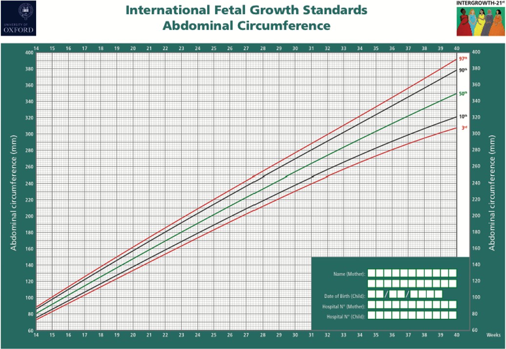
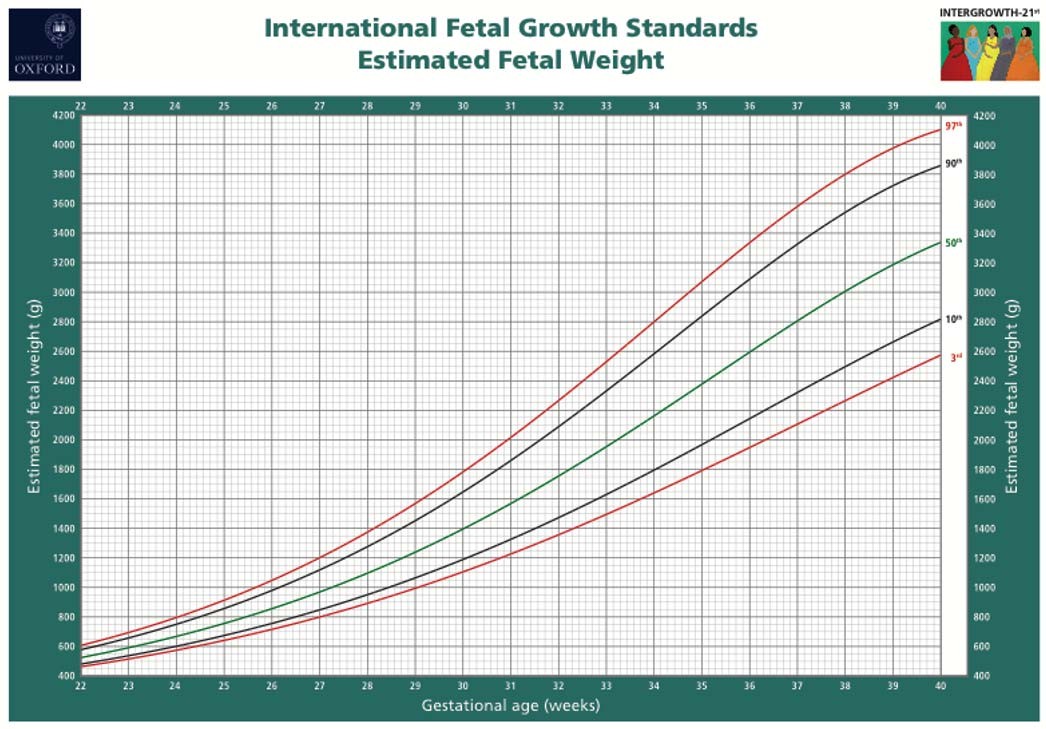
The Intergrowth-21st universal chart is attached in this guideline (Appendix 2).
What further investigations should be performed when FGR is suspected?
Once FGR is suspected, a systematic investigation should be performed to identify the underlying aetiology, the most important reasons being consti-tutional SGA, placental dysfunction, and fetal conditions such as genetic or infective disorders. The investigation should include a detailed history, evaluation of screening test results for trisomy 21 and biochemical markers, detailed anatomy scan to assess structural anomalies, Doppler studies and additional testing directed at genetic or infectious aetiologies when suspected.
Detailed history
A detailed maternal and family history is essential to identify the aetiology correctly. It should include maternal age, racial or ethnic group, height and weight, nutritional status, socio-economic status, medications, cigarette smoking, use of recreational drugs, chronic medical conditions, personal or family history of thrombophilia, genetic disorders or consanguinity, obstetrics history including the birth weight of previous children and confirmation of pregnancy dating by first-trimester ultrasound.
A history of febrile illness or rash in pregnancy or periconceptional period, recent travel history to endemic areas and frequent exposure to young children or domestic animals should be obtained to assess the risk of congenital fetal infection with cytomegalovirus, toxoplasmosis, rubella, syphilis, zika virus and varicella zoster virus.
Accurate dating of pregnancy is essential for correctly interpreting estimated fetal size and avoiding a false diagnosis of FGR. Except for pregnancies achieved by assisted reproductive technology, the crown-rump length (CRL) measured at the time of 1st trimester ultrasound when in the range of 7-60mm is the most accurate method to date the pregnancy and establishes gestational age with the precision of 5 days in 95% cases. If more than one scan is performed in the 1st trimester, the earliest scan with a CRL of at least 10mm should be used1.
Detailed anatomy scan
A detailed anatomy scan should be performed when FGR is suspected, especially in early-onset FGR.
The presence of major structural anomalies, soft markers or polyhydramnios may raise the possibility of chromosomal, sub-chromosomal, or single-gene abnormalities. The presence of shortened fetal long bones (shorter than -2SD, especially -4SD below the mean) should raise the possibility of skeletal dysplasia and indicates targeted genetic assessment. The sonographic findings of small head circumference, ventriculomegaly, brain or liver calcifications, periven-tricular hyperechogenicity, cortical brain malfor-mations, echogenic bowel, hydrops or placentomegaly should raise the suspicion of congenital infections, especially in women with relevant history.
Doppler studies
Doppler assessment is integral to the diagnosis, surveillance, and management of FGR.
In all cases of suspected FGR, serial monitoring of umbilical artery Doppler studies is recommended, as it may be normal in the early stages of placental FGR. Abnormal umbilical artery Doppler is not patho-gnomonic of placental dysfunction as certain genetic conditions such as triploidy may also mimic early-onset placental FGR with abnormal umbilical artery Doppler secondary to abnormal placental karyotype.
Uterine artery Doppler is less likely to be abnormal among fetuses with FGR and abnormal karyotype and therefore considered more specific for primary placental FGR, especially in the presence of abnormal angiogenic markers in maternal blood.
Additional testing
- Screening for congenital infections
Should be offered when FGR is suspected, especially in early-onset FGR or when an infection is possible based on history or ultrasound findings. Testing should include CMV and toxoplasmosis but may also include rubella, varicella, and syphilis in women at increased risk for these infections. Women with recent travel history to endemic areas should be offered testing for Zika virus and malaria.
When fetal infection is highly suspected based on serology or clinical findings, amniocentesis should be offered where expertise and resources are available, to detect viral DNA in the amniotic fluid using poly-merase chain reaction (PCR). Amniocentesis should be delayed until after 21 weeks’ gestation and at least 6-8 weeks following the estimated onset of maternal infection to minimize the risk of false negative results.
Genetic testing
Referral to a geneticist and genetic testing by amniocentesis should be offered to women with FGR, especially early-onset or severe FGR (<3rd centile), co-presence of sonographic findings (structural anomalies, soft markers, or polyhydramnios) and the absence of apparent signs of placental dysfunction such as abnormal uterine or umbilical artery Doppler.
Women should be counselled about the risk of a genetic aetiology even in isolated FGR without associated fetal anomalies. It is reasonable to offer amniocentesis with karyotype and microarray analysis, when resources and expertise available, to women with FGR with the decision based on factors such as ultrasound findings, gestational age, lack of evidence of placental dysfunction and if the results of amniocentesis would affect management.
Management of FGR / SGA
Monitoring
The primary goal of fetal monitoring is the prevention of stillbirth by detecting fetal deterioration that precedes irreversible compromise.
Fetal surveillance tests include;
- Fetal movement counting
- Fetal heart rate (FHR) monitoring with cardiotocography (CTG)
- Computerized CTG and short-term variation
- Ultrasound evaluation of amniotic fluid volume
- Biophysical profile (BPP) scoring
- Doppler velocimetry of umbilical artery, middle cerebral artery, and ductus venosus
Fetal movement counting
Decreased fetal movement is defined as less than 10 movements in 2 hours during focused maternal counting when lying down10. It reflects the reduction in fetal activity due to progressive fetal hypoxemia. It is a simple and inexpensive tool that may provide a safety net between scheduled outpatient monitoring visits and can be used as an adjunct to the monitoring of FGR.
- Fetal heart rate (FHR) monitoring with CTG
Antepartum CTG, also known as nonstress test (NST), can be performed alone or in conjunction with the measurement of amniotic fluid volume known as a modified biophysical profile (BPP) or a five-component BPP. FHR pattern reflects fetal oxygenation and acid-base status at the time of evaluation but does not predict deterioration in FGR. A nonreactive CTG/NST has low specificity for hypoxia and requires additional tests to determine fetal status and distinguish FHR pattern variation caused by fetal behaviour, while reduced variability is a much stronger predictor of central nervous system hypoxia.
The empirically recommended frequency of CTG in FGR is twice weekly, with the frequency increasing when the evaluation of amniotic fluid or Doppler parameters indicates a more advanced degree of fetal compromise.
Computerized CTG and short-term variation (STV)
Unlike conventional CTG, which has high intra- and inter-observer variation, computerized CTG is objective and consistent. Fetal heart variability is the most useful predictor of fetal wellbeing in SGA fetuses. An STV
3 ms (within 24 hours of delivery) has been associated with a higher rate of metabolic acidaemia and early neonatal death.
After 29 weeks’ gestation, below 4.0 ms or 3.0 ms meet the criteria for reduced or very low STV, respectively. Before 29 weeks’ gestation, STV below
3.5 ms is considered reduced, and below 2.6 ms is considered very low.
cCTG does not predict fetal deterioration, like CTG/ NST. Monitoring with CTG/NST or cCTG needs to be performed more frequently than Doppler studies, and among those receiving inpatient monitoring, a minimum frequency of daily CTG/NST or cCTG is recommended.
Ultrasound evaluation of amniotic fluid volume
A decrease in amniotic fluid volume can result from fetal oliguria in response to progressive placental dysfunction and hypoxia, as well as rupture of membranes. Oligohydramnios is defined as the amniotic fluid index (AFI) 5 cm or deepest vertical amniotic fluid pocket (DVP) 2 cm. DVP is preferred over AFI as it reduces the overdiagnosis of oligo-hydramnios.
Oligohydramnios is associated with increased intrapartum FHR abnormalities, need for caesarean section and Apgar scores of less than 5, but not acidosis at birth. Assessment of amniotic fluid volume is not recommended in isolation into the management decision of FGR.
Biophysical profile (BPP) scoring
BPP scoring is not recommended as the primary surveillance tool for FGR. The five-component BPP score consists of fetal breathing movements, gross body movements, fetal tone, maximum amniotic fluid pocket and CTG. The modified BPP refers to the combined use of the CTG/NST as a short-term indicator of acid-base balance and the maximum amniotic fluid pocket as an indicator of long-term placental function.
In the five-component BPP, a score of 4 is considered abnormal. The modified BPP is considered abnormal when either the CTG/NST is nonreactive, or the maximum amniotic fluid pocket is <2cm. A BPP score of 4 is associated with a fetal pH 7.20, while a score of <2 has a sensitivity of 100% for acidemia. This correlation remains highly significant even when using modified BPP11.
Doppler velocimetry of umbilical artery, middle cerebral artery, and ductus venosus
- Umbilical artery Doppler
In the high-risk population, UA Doppler has been shown to reduce perinatal morbidity and mortality and should be the primary surveillance tool in the SGA fetus.
It is estimated that approximately one-third of the villous circulation must be damaged before a decrease in UA EDF velocity occurs, while absent or reversed UA EDF corresponds to the malperfusion of 50-70% of the villous vascular tree. As the elevated villous blood flow resistance is predominantly associated with placental pathology seen in early-onset FGR, UA Doppler does not reliably predict outcomes in late-onset FGR.
When UA Doppler flow indices are normal, it is reasonable to repeat surveillance every 2 weeks. Where UA PI is > 95th centile with the presence of EDF, it requires a surveillance frequency of once or twice a week. When AEDF develops, Doppler surveillance is recommended at minimum twice weekly and for REDF at least three times a week unless delivery criteria has been met1.
Middle cerebral artery Doppler
It is recommended to use MCA Doppler for monitoring late-onset FGR. Concurrent measurement of UA and the MCA pulsatility index allows calculation of the cerebro-placental Doppler ratio (CPR). CPR and MCA-PI decrease as a hemodynamic response to fetal hypoxemia and reflect placental dysfunction, even in pregnancies where the villous blood flow resistance is not elevated enough to produce an abnormal UA-PI.
Approximately 20% of term SGA fetuses with normal UA Doppler have a decreased MCA-PI, which is associated with a higher caesarean section rate due to fetal distress or poor neonatal transition and adverse developmental outcomes. Therefore, an important role of MCA Doppler is to estimate perinatal risk in patients with normal UA Doppler.
Because of the higher risk for adverse outcomes within one week of a decrease in MCA-PI, it is recommended to utilize at least twice weekly surveillance1.
Ductus venosus Doppler
Abnormal ductus venosus Doppler is primarily observed in early-onset FGR and can estimate fetal acid-base balance and the risk of stillbirth. The relative forward flow in atrial systole in the ductus venosus decreases with worsening placental function or reduced fetal cardiac function leading to an increase in PI of veins, absence, or reversal of a-wave.
In fetuses with elevated ductus venosus PI of veins but forward flow during atrial systole, the median interval to progressive venous Doppler deterioration can be as short as two days. Therefore, in patients that do not meet the delivery criteria, ductus venous Doppler is recommended at minimum twice weekly in those with UA Doppler showing AEDV and thrice weekly when REDV is observed. When the ductus venous Doppler indices increase as a new finding, the frequency of monitoring needs to be increased further.
Timing of delivery
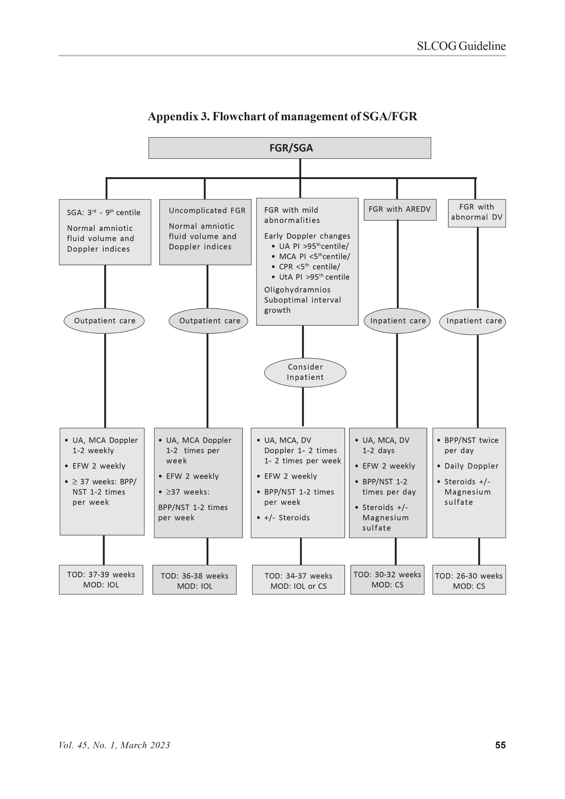
Timing of delivery in FGR is determined by gestational age, the severity of FGR, findings of fetal monitoring tests and maternal factors such as pre-eclampsia. Recommendations for monitoring, timing, and mode of delivery in SGA are summarized in Table 5 and Appendix 3.
Gestational age-related risks in FGR
Between 24-28 weeks of gestation, each day of pregnancy prolongation results in an estimated 2% decrease in perinatal morbidity and mortality. Between 28-30 weeks of gestation, the daily increment in survival is 0.7%. After 30 weeks, the neonatal survival rate exceeds 90%. From 34-38 weeks of gestation, neonates are more likely to require admission to the intensive care units but have reduced risks of significant perinatal morbidities.
SGA fetuses undelivered after 38 weeks of gestation, the risk of stillbirth doubles every week and reaches 6/1000 for pregnancies that continue beyond the due date.
Absolute criteria requiring delivery (independent of gestational age)
A 30-minute BPP score of 0 or 2 indicates prelabour fetal pH of less than 7.20 and requires delivery to prevent fetal demise.
- In conventional CTG, repetitive fetal heart rate decelerations, sinusoidal heart rate, and absent variability with recurrent late decelerations or fetal bradycardia predict fetal academia and poor perinatal outcome and require delivery if the causative stimulus cannot be removed.
- When computerized CTG is used, a short-term variation below 2.6 ms is below the 5th percentile irrespective of gestational age and requires delivery for its strong association with fetal academia.
- Pre-eclampsia with uncontrolled hypertension, HELLP syndrome or other evidence of end-organ damage requires delivery.
Management of early onset FGR
Once early FGR is suspected or diagnosed, the pregnancy should be monitored and managed in the tertiary-level unit where neonatal facilities are available. Multidisciplinary counselling by a neonatologist and consultant Obstetrician is essential. The surveillance frequency should be based on the severity of FGR and umbilical artery abnormalities.
It is recommended to monitor the EFW and Doppler indices two weekly if the UA Doppler is normal. If the UA-PI >95th centile, twice weekly Doppler with fortnightly EFW is recommended. Progressive deterioration of UA Doppler (AEDV or REDV) warrants more intensive monitoring every 2-3 days with fortnightly EFW.
MCA Doppler is one of the first parameters that become abnormal in early FGR. There seems to be a weak association between low MCA-PI and adverse neonatal outcomes. There is no evidence that it should be used to determine delivery timing.
Monitoring and delivery timing based on the ductus venous Doppler and computerized CTG provides better outcomes (TRUFFLE trial)12. Absent or reversed a-wave in the ductus venosus Doppler or computerized CTG- short-term variation <3ms warrants delivery. If computerized CTG is unavailable, it is recommended to use conventional CTG and BPP scoring in addition to the Doppler indices.
Around 70% of early FGR is associated with hypertensive disorders of pregnancy, mainly pre-eclampsia. Thus, regular blood pressure assessment, monitoring of urine protein/creatinine ratio and baseline renal and hepatic function is recommended for asymptomatic women with early FGR.
Management of Late-onset FGR
Alterations in UA Doppler and DV Doppler are rare and fail to identify the vast majority of late-FGR or to predict adverse outcomes.
A reduction in MCA-PI or its ratio with UA-PI (CPR) is associated with poorer perinatal outcomes, including stillbirth and a higher risk of caesarean delivery. CPR can identify subtle changes between placental and cerebral blood-flow perfusion that may not be appreciated by evaluating a single parameter and may improve the prediction of adverse perinatal outcomes in growth-restricted fetuses.
BPP abnormalities that characterize late FGR include alteration of fetal breathing, decreased amniotic fluid volume and loss of fetal heart rate reactivity on conventional CTG. In late FGR, it seems that BPP becomes abnormal only shortly before stillbirth; therefore, it does not help determine monitoring intervals.
Table 5. Summary of recommendations for monitoring, timing, and mode of delivery
| Findings | Risk of stillbirth | Monitoring | Time and mode of delivery |
|---|---|---|---|
| SGA: EFW at 3rd to 9th percentile, normal amniotic fluid volume and normal Doppler indices | Low | UA, MCA Doppler every 1-2 weeks EFW fortnightly At 37 weeks: consider BPP/NST 1-2 times per week | 37-39 weeks Induction |
| Uncomplicated FGR: EFW < 3rd centile with normal amniotic volume and normal Doppler indices | Low | UA, MCA Doppler 1-2 times per week EFW fortnightly At 37 weeks: consider BPP/NST 1-2 times per week | 36-38 weeks Induction |
| FGR with mild abnormalities: Early Doppler changes UA PI >95th percentile, or MCA PI < 5th percentile, or CPR < 5th percentile, or UtA PI>95th percentile Oligohydramnios Suboptimal interval growth Suspected pre-eclampsia | Low | Consider inpatient monitoring Offer steroids for fetal lung maturation between 24+0 to 34+6 weeks BPP/NST 1-2 times per week UA, MCA, DV Doppler 1-2 times per week EFW fortnightly | 34-37 weeks Caesarean section or induction |
| FGR withAEDV/REDV Median time of deterioration: AEDV: 5 days REDV: 2 days | AEDV-6.8% REDV-19% | Inpatient monitoring Offer steroids for fetal lung maturation between 24+0 to 34+6 weeks Offer magnesium sulfate for neuroprotection between 24+0 to 29+6 weeks and considered between 30+0 to 33+6 weeks BPP/NST: 1-2 times per day UA, MCA, DV Doppler: 1-2 days EFW fortnightly | 30-32 weeks Caesarean section |
| FGR with abnormal Ductus Venosus Doppler | 20% | Inpatient monitoring Offer steroids for fetal lung maturation between 24+0 to 34+6 weeks Offer magnesium sulfate for neuroprotection between 24+0 to 29+6 weeks and considered between 30+0 to 33+6 weeks BPP/ NST twice per day Daily Doppler | 26-30 weeks Caesarean section |
In the presence of UA-PI>95th centile, monitoring of MCA-PI and UA-PI is indicated at least once or twice a week.
In pregnancies with late FGR and UA-PI >95th percentile, delivery should be considered at 36+0 weeks and not later than 37+6 weeks of gestation. When there is late FGR with signs of cerebrovascular redistri-bution, it is recommended to deliver by 38+0 weeks and no later than 38+6 weeks of gestation11.
Management of small-for-gestational-age
SGA categorization is frequently applied to a small baby that is structurally normal and has normal Doppler findings. Adopting customized growth charts has been suggested to reduce the proportion of SGA.
Evidence suggests that SGA with normal fetoplacental Doppler can be associated with accelerated placental ageing, signs of placental under perfusion, lower umbilical vein blood flow volume, altered maternal hemodynamics and greater incidence of caesarean section for fetal distress compared with average gestational age fetuses.
At the diagnosis of SGA, fetal Doppler indices (UA PI, MCA PI, and CPR) and uterine artery Doppler should be evaluated. Thereafter, fortnightly assessment of fetal growth and weekly assessment of Doppler indices (UA PI, MCA PI, CPR) is recommended.
Once SGA is identified, it is recommended to deliver from 38+0 weeks gestation, not exceeding 39+0 weeks (DIGITAT study)13,14.
Mode of delivery and intrapartum considerations
FGR itself is not an indication for caesarean section. Decisions on the mode of delivery should be based on the gestational age, the severity of FGR, Doppler changes, associated maternal co-morbidities such as pre-eclampsia, parity, cervical Bishop score and patient preference.
In severe early onset FGR, the main goal is to prolong pregnancy and maximize fetal maturation by conservative management with close surveillance until evidence of Doppler changes. As the fetus might already be experiencing some degree of hypoxia or acidosis, the likelihood of tolerating labour is low, with reported caesarean section rates of >80%. Also, labour induction is less likely to succeed during the preterm period. For these reasons, primary caesarean section is usually the preferred option for severe early-onset FGR.
Late-onset FGR is usually less severe, and fetal hypoxia or acidosis is less likely to be present when delivery is indicated. Therefore, labour induction can be considered for most late-onset FGR in the presence of normal UA Doppler and the absence of additional factors.
The optimal method for cervical ripening for FGR is unclear. Mechanical methods (balloon catheter) appear to be associated with a lower risk of caesarean section and intrapartum complications than prostaglandin induction. If prostaglandin is used for induction, a reversible method (e.g., dinoprostone vaginal insert) is preferable.
During labour, continuous fetal monitoring is recommended. Delivery should occur at an institution with the appropriate level of neonatal care for the gestational age and the anticipated management needs of the neonate.
It is recommended to send the placenta for histopathological examination after delivery. High-quality evaluation of the placental pathology is likely to increase the precision of diagnosis and provide information on the risk of recurrence.
Medical interventions
- Antenatal corticosteroids
A course of antenatal corticosteroids should be offered between 24+0 to 34+6 weeks to women whose babies are thought to be either SGA or to have FGR to reduce neonatal morbidity and mortality15. Enhanced daily surveillance is warranted during steroid administration in fetuses with absent or reversed UA-EDF.
No RCTs have been performed to establish if the benefit of corticosteroid prophylaxis in premature fetuses also applies to premature growth-restricted fetuses in whom the reduced metabolism of corticosteroids by a small placenta and already high levels of endogenous adrenal corticosteroids might further damage the brain’s white matter and myelination16.
Magnesium sulphate for neuroprotection
Administration of magnesium sulphate to women at risk of preterm birth has been shown to have a neuroprotective role, with a decrease in the risk of perinatal mortality, cerebral palsy and gross motor dysfunction.
Intravenous magnesium sulphate for neuroprotection for preterm fetuses is offered between 24+0 to 29+6 weeks of gestation and should be considered between 30+0 to 33+6 weeks of gestation17. Magnesium sulphate should be administered as a 4 grams bolus intravenously over 15 minutes, followed by an intravenous infusion of 1 gram per hour until the birth or for 24 hours, whichever is sooner17.
Postpartum assessment and counselling for future pregnancies
Infant follow-up
Growth-restricted infants are at increased risk of immediate and long-term complications and therefore require closer follow-up than normally grown infants in the first years of life.
Maternal follow-up
Women with a history of FGR or other placenta-mediated complications such as pre-eclampsia require follow-up as they are at an increased risk of future cardiovascular disease, especially in early-onset disease.
Counselling regarding future pregnancies
- Risk of recurrence based on severity and onset
In women with a previous FGR fetus <10th centile, the chance of recurrence in subsequent pregnancies is less than 25%. The recurrence risk increases with the severity of FGR and is nearly 6-fold when the infant’s birth weight is below the 5th centile.
Counselling regarding the risk of recurrence should be refined based on the individual patient’s risk factors, the severity of FGR as reflected by the timing of onset and Doppler findings, the co-presence of pre-eclampsia, and placental histopathological findings.
- Risk of recurrence based on placental histopathology
The results of the placental histopathological examination are important for two main reasons: First, they may assist in counselling couples regarding the most likely aetiology of FGR, especially when the clinical presentation and Doppler findings were inconclusive. Second, placental findings may provide valuable information regarding the risk of recurrence, as certain placental pathologies, such as villitis of unknown aetiology and chronic histiocytic intervillositis, are associated with relatively high recurrence of 10-50% and 70-100%, respectively.
Role of thrombophilia screening
There is insufficient evidence to justify routine screening for antiphospholipid (aPL) antibodies in women with prior FGR.
Antiphospholipid syndrome (APLS) screening is, however, recommended in women with a history of thromboembolism or recurrent pregnancy loss or 1 late fetal loss and may be considered in selected cases of women with a history of severe FGR associated with severe early-onset pre-eclampsia and when placental examination shows features of severe maternal vascular malperfusion.
Management of women already diagnosed with APLS based on a history of placenta-mediated complications is debatable. Based on the available evidence FIGO guidelines recommend treatment with aspirin, and suggest LMWH be considered only in selected cases, such as for women who have experienced recurrent complications despite aspirin treatment (aspirin failure)1.
Preconception counselling and management of future pregnancies
Given the considerable risk of recurrence of FGR, efforts should be made to decrease this risk in future pregnancies. Modifiable risk factors for FGR, such as smoking or poor nutritional status, should be identified as early as possible and managed accordingly.
Aspirin should be considered in women with past FGR only if they have risk factors for pre-eclampsia at the time of the next pregnancy. However, FIGO guidelines suggest that given the safety of aspirin and the overlap in the pathogenesis of pre-eclampsia and FGR, it is reasonable to recommend aspirin to women with a history of placenta mediated FGR in the previous pregnancy, using the same regimen of aspirin used for prevention of pre-eclampsia. LMWH therapy should not be used in women with a history of FGR except in a research setting.
Given the association of insufficient gestational weight gain with FGR, it is recommended to provide information about the target weight gain and monitor weight gain. Other interventions, such as bed rest or nutritional supplements, are of unproven benefit and should not be routinely offered.
Due to the increased risk of recurrence, pregnant women with a history of FGR in a previous pregnancy should receive closer antenatal surveillance, including close monitoring of fetal growth and maternal blood pressure.
Appendix 2. Intergrowth-21st chart for EFW and AC
References
- Melamed N, Baschat A, Yinon Y, Athanasiadis A, Mecacci F, Figueras F, et al. FIGO (International Federation of Gynecology and Obstetrics) initiative on fetal growth: best practice advice for screening, diagnosis, and management of fetal growth restriction. Int J Gynaecol Obstet. 2021; 152 Suppl 1(Suppl 1): 3-57.
- Royal College of Obstetricians and Gynaecologists GtGN. The Investigation and Management of the Small-for-Gestational-Age Fetus. 2nd Edition edFebruary 2013 | Minor revisions - January 2014.
- Roberge S, Nicolaides K, Demers S, Hyett J, Chaillet N, Bujold E. The role of aspirin dose on the prevention of preeclampsia and fetal growth restriction: systematic review and meta-analysis. Am J Obstet Gynecol. 2017; 216(2): 110-20.e6.
- Meher S, Duley L. Progesterone for preventing pre-eclampsia and its complications. Cochrane Database Syst Rev. 2006; 2006(4): CD006175.
- Hofmeyr GJ, Lawrie TA, Atallah Á, Torloni MR. Calcium supplementation during pregnancy for preventing hypertensive disorders and related problems. Cochrane Database Syst Rev. 2018; 10(10): CD001059.
- Rodger MA, Gris JC, de Vries JIP, Martinelli I, Rey É, Schleussner E, et al. Low-molecular-weight heparin and recurrent placenta-mediated pregnancy complications: a meta-analysis of individual patient data from randomised controlled trials. Lancet. 2016; 388(10060): 2629-41.
- Shamawarna K, Goonewardene M, Perera Y. Customised symphysio fundal height charts. The Ceylon Medical Journal. 2012; 57: 159-65.
- Milner J, Arezina J. The accuracy of ultrasound estimation of fetal weight in comparison to birth weight: A systematic review. Ultrasound. 2018; 26(1): 32-41.
- Sovio U, White IR, Dacey A, Pasupathy D, Smith GCS. Screening for fetal growth restriction with universal third trimester ultrasonography in nulliparous women in the Pregnancy Outcome Prediction (POP) study: a prospective cohort study. Lancet. 2015; 386(10008): 2089-97.
- Moore TR, Piacquadio K. A prospective evaluation of fetal movement screening to reduce the incidence of antepartum fetal death. Am J Obstet Gynecol. 1989; 160(5 Pt 1): 1075-80.
- Lees CC, Stampalija T, Baschat A, da Silva Costa F, Ferrazzi E, Figueras F, et al. ISUOG Practice Guidelines: diagnosis and management of small-for-gestational-age fetus and fetal growth restriction. Ultrasound Obstet Gynecol. 2020; 56(2): 298-312.
- Bilardo CM, Hecher K, Visser GHA, Papageorghiou AT, Marlow N, Thilaganathan B, et al. Severe fetal growth restriction at 26-32 weeks: key messages from the TRUFFLE study. Ultrasound Obstet Gynecol. 2017; 50(3): 285-90.
- Boers KE, Vijgen SMC, Bijlenga D, van der Post JAM, Bekedam DJ, Kwee A, et al. Induction versus expectant monitoring for intrauterine growth restriction at term: randomised equivalence trial (DIGITAT). BMJ. 2010; 341: c7087.
- Boers KE, van Wyk L, van der Post JA, Kwee A, van Pampus MG, Spaanderdam ME, et al. Neonatal morbidity after induction vs expectant monitoring in intrauterine growth restriction at term: a subanalysis of the DIGITAT RCT. Am J Obstet Gynecol. 2012; 206(4): 344.e1-7.
- Stock S, Thomson A, Papworth S, Gynaecologists tRCoOa. Antenatal corticosteroids to reduce neonatal morbidity and mortality. BJOG: An International Journal of Obstetrics and Gynaecology. 2022; 129(8): e35-e60.
- Magann EF, Haram K, Ounpraseuth S, Mortensen JH, Spencer HJ, Morrison JC. Use of antenatal corticosteroids in special circumstances: a com-prehensive review. Acta Obstetricia et Gynecologica Scandinavica. 2017; 96(4): 395-409.
- guideline N. Preterm labour and birth. Last updated 10 June 2022 ed 20 November 2015.