
Background
Shoulder dystocia is a serious obstetric emergency as far as the fetus is concerned. It occurs as a result of the fetal anterior shoulder being impacted above the maternal pubic symphysis, while the fetal posterior shoulder would have descended below the maternal sacral promontory. The anatomical relationships of the lumbar vertebrae, sacral promontory and the sacral hollow almost always enable the posterior shoulder to slip underneath the sacral promontory, and therefore, bilateral shoulder dystocia, where both fetal shoulders are above the pelvic brim, is only a theoretical possibility. Although the global incidence of shoulder dystocia is quoted as approximately 1%, inconsistency in the reporting and controversies surrounding the diagnosis make an accurate determination of its incidence difficult. There is no data specific to Sri Lanka.
Summary of recommendations
- An experienced practitioner (Consultant, Senior Registrar, Registrar or Senior House Officer) should be present at delivery when any of the risk factors for shoulder dystocia are present.
- As shoulder dystocia is largely unpredictable, every birth attendant should be conversant with the diagnosis, and at least, the initial management of shoulder dystocia.
A diagnosis of shoulder dystocia should be made;
- if the fetal shoulders fail to deliver spontaneously or with routine axial traction on the head OR if the fetal chin is tightly pressed against the perineum (turtle sign).
- Once shoulder dystocia occurs, effort should be made to complete the delivery of the fetus within four minutes, provided that the fetus was not compromised before the event.
- Measures that would aggravate the shoulder dystocia or that may cause injury, such as excessive down- ward traction on the fetal head, fundal pressure or maternal pushing should be avoided.
- Once shoulder dystocia is diagnosed, the most experienced practitioner on site, the neonatal team, and additional personnel to place the woman in positions that may be required for delivery, should be called for. Brief explanation about the situation should be provided to the woman.
- Mild, axial traction, only sufficient to help the fetal anterior shoulder to slip underneath the maternal pubic symphysis, should be applied following each manoeuvre that is attempted to manage the shoulder dystocia.
- If one manoeuvre is not successful, one should not persist with the same ineffective manoeuvre for long and should adopt the next most feasible manoeuvre within a reasonable time frame of approximately 30 seconds.
- The woman should be taken to the lower edge of the bed, or the lower half of the bed should be dismantled, to facilitate the application of traction on the fetal head in the desired direction.
- As a first-line measure, the woman should be placed in McRobert’s position, with simple instructions to the assistants.
- If McRobert’s manoeuvre fails, suprapubic pressure should be applied in a downward and lateral direction, ideally from the direction of the posterior aspect of the fetal anterior shoulder, with the heels of both hands, either continuously or by rocking movements.
- If first-line measures fail, then delivery of the posterior arm or direct rotation of the fetal shoulders should be attempted, with the adoption of measures that would maximize the chances of safe entry into the vagina as well as effective delivery of the shoulders. All-fours manoeuvre could be considered if feasible.
- One could attempt to deliver the posterior arm by sweeping it across the fetal chest and face or by applying traction on the posterior axilla, depending on personal preference and the severity of the shoulder dystocia.
- If the shoulder dystocia remains unresolved even after the posterior arm is delivered, one should attempt to rotate the fetus by 180° and complete the delivery.
- One should attempt to rotate the fetal shoulders to an oblique diameter of the pelvis or by 180° by the application of direct pressure on one or both shoulders, using one or a set of manoeuvres in a logical sequence, with suprapubic pressure in a complementary direction.
- Placing the woman in all-fours position could be considered as a second-line measure.
- Third-line manoeuvres are seldom indicated in the management of shoulder dystocia, and if required, should be performed by a team led by an experienced obstetrician.
- Following shoulder dystocia, the baby should be handed over to the neonatal team, and the woman should be examined for bleeding as well as genital tract and perineal trauma.
- Accurate documentation, debriefing of the woman, her partner and staff, risk management and regular training are pivotal for governance issues surrounding shoulder dystocia.
- Elective caesarean delivery if the estimated fetal weight is greater than the 95th centile at term for a given population or with previous history of permanent brachial plexus injury following shoulder dystocia, and induction at 37-38 weeks of gestation in both diabetic and non-diabetic women with suspected fetal macrosomia should be considered as preventive measures for shoulder dystocia.
- There is a need for a strong predictive system and a universally acceptable definition for shoulder dystocia and recognition of the primary clinicians’ role in the management of shoulder dystocia.
Risk factors and prediction
The risk factors for shoulder dystocia are given in Table 1.
Table 1. The risk factors for shoulder dystocia
| Maternal characteristics | Maternal obesity Excessive maternal weight gain during pregnancy |
|---|---|
| Previous obstetric history | Previous shoulder dystocia |
| Antepartum | Fetal macrosomia Maternal diabetes mellitus Post-term pregnancy |
| Intrapartum | Augmented labour Prolonged first stage of labour Prolonged second stage of labour Instrumental vaginal delivery |
Despite the low positive predictive value of these risk factors, anticipation is key to early diagnosis and prompt management of shoulder dystocia.
An experienced practitioner (Consultant, Senior Registrar, Registrar or Senior House Officer) should be present at delivery when any of the above risk factors for shoulder dystocia are present.
However, shoulder dystocia can occur in any delivery, and for all practical purposes, it is neither predictable nor preventable.
Therefore, every birth attendant should be conversant with the diagnosis, and at least, the initial management of shoulder dystocia.
Diagnosis of shoulder dystocia
Usually, the fetal shoulders deliver either spontaneously or with gentle traction on the head in the direction of the longitudinal axis of the fetus (routine axial traction).
A diagnosis of shoulder dystocia should be made;
- if the fetal shoulders fail to deliver spontaneously or with routine axial traction on the head OR
- if the fetal chin is tightly pressed against the perineum (turtle sign)
The need for additional manoeuvres to deliver the fetal shoulders, although useful as an audit tool in retrospect, is not helpful for the purposes of early diagnosis and management of shoulder dystocia.
Management of shoulder dystocia
Time limits
Once shoulder dystocia occurs, the ongoing uterine contractions lead to a reduction of blood flow to the intervillous space, and furthermore, the fetal chest is unable to expand due to compression within the maternal pelvis. As a result, birth asphyxia can occur within a short period of time, with possible permanent brain damage if not delivered within 4-5 minutes.
Once shoulder dystocia occurs, effort should be made to complete the delivery of the fetus within four minutes.
This 4 minute leeway is not acceptable if the fetus is already compromised. Nevertheless, one should refrain from exercising undue haste, which has the potential to cause birth trauma and enhance the adverse effects of hypoxia.
What to avoid
Persisting with the application of excessive downward traction on the fetal head is one of the main reasons for brachial plexus injury, and this should be avoided. Similarly, fundal pressure (which also has the potential to cause uterine rupture) or maternal pushing, which aggravates the shoulder dystocia, should be discouraged.
Measures that would aggravate the shoulder dystocia or that may cause injury, such as excessive downward traction on the fetal head, fundal pressure or maternal pushing should be avoided.
Communication
Once shoulder dystocia is diagnosed, the most experienced practitioner on site, the neonatal team, and additional personnel to place the woman in positions that may be required for delivery, should be called for. Brief explanation about the situation should be provided to the woman.
It is good practice to allocate a staff member to keep track of time and events.
Manoeuvres
Shoulder dystocia should be managed with any of the manoeuvres mentioned below.
Mild, axial traction, only sufficient to help the fetal anterior shoulder to slip underneath the maternal pubic symphysis, should be applied following each manoeuvre that is attempted to manage the shoulder dystocia.
If one manoeuvre is not successful, one should not persist with the same ineffective manoeuvre for long and should adopt the next most feasible manoeuvre within a reasonable time frame of approximately 30 seconds.
First-line measures
It is important that traction in the direction of the longitudinal axis of the fetus (axial traction) is applied on the fetal head after each manoeuvre, as traction in any other direction can cause injuries to the fetal cervical spine. This would mean a slightly downward direction, at an angle of approximately 30° to the horizontal.
The woman should be taken to the lower edge of the bed, or the lower half of the bed should be dismantled, to facilitate the application of traction on the fetal head in the desired direction.
First-line measures: McRobert’s position
McRobert’s position, i.e. hyperflexion of thighs with knees brought towards the woman’s chest with the help of two assistants, leads to a slightly increased anteroposterior diameter of the maternal pelvis, but more importantly, brings the longitudinal axis of the pelvis in line with the direction of forces required for delivery, thereby making these forces more effective. As a first-line measure, the woman should be placed in McRobert’s position, with simple instructions to the assistants.
The most experienced practitioner attending the delivery should position him/herself between the maternal thighs and apply mild, axial traction on the fetal head.
First-line measures: Suprapubic pressure
If the shoulder dystocia is not resolved by McRobert’s manoeuvre, the woman’s thighs should be abducted to allow a third assistant to apply suprapubic pressure. If a third assistant is not available, the one who is performing McRobert’s manoeuvre should apply suprapubic pressure. The main aim of suprapubic pressure is to rotate the fetal shoulders which are stuck in the anteroposterior diameter to the wider oblique diameter of the pelvic inlet.
Application of suprapubic pressure from the posterior aspect of the anterior shoulder has the additional advantage of causing adduction of that shoulder, which would lead to a reduction of the effective diameter of the shoulder girdle. The direction of the fetal face, if restitution has taken place, or the previous examination findings can be used as a guide to the desired direction of suprapubic pressure. Suprapubic pressure should be applied in a downward and lateral direction with the heels of both hands, one on top of the other with fingers interlocking (see Figure 1 in annexure). Continuous pressure or rocking movements are equally effective.
If McRobert’s manoeuvre fails, suprapubic pressure should be applied in a downward and lateral direction, ideally from the direction of the posterior aspect of the fetal anterior shoulder, with the heels of both hands, either continuously or by rocking movements.
Second-line manoeuvres
In case of failure of the first-line measures, then second-line manoeuvres, which involve delivery of the posterior arm or direct rotation of the fetal shoulders, should be adopted. Either method is acceptable depending on the preference of the obstetrician and the feasibility according to the clinical circumstances. Upon inserting the operator’s hand into the vagina, if the posterior arm is flexed, then delivery of the posterior arm, and if the arm is extended, then direct rotation of the shoulder/s, is justifiable.
Although an episiotomy does not correct the shoulder dystocia, it could be considered, or the existing episiotomy could be extended, in order to improve access into the vagina. When gaining access into the vagina, the operator’s whole hand should be inserted, initially, with the thumb and fingers held together in the shape of a cone (see Figure 2 in annexure). This would prevent injury to maternal tissues.
As the anterior aspect of the vagina is filled with the body of the fetus, the operator should initially insert the hand/s into the posterior aspect of the vagina, even if a manoeuvre involves application of direct pressure on the anterior shoulder later.
If first-line measures fail, then delivery of the posterior arm or direct rotation of the fetal shoulders should be attempted, with the adoption of measures that would maximize the chances of safe entry into the vagina as well as effective delivery of the shoulders. All-fours manoeuvre could be considered if feasible.
Second-line manoeuvres: Delivery of the posterior arm
The posterior arm can be delivered by either sweeping it across the fetal chest and face or by applying traction on the posterior axilla, which is gaining popularity as an easier, more effective, and less traumatic method.
When attempting to deliver the posterior arm across the fetal chest, the wrist should be grasped between the fingers and the thumb of the operator, and traction should be applied in a straight line downwards in line with the forearm (see Figure 3 in annexure). If the posterior hand and forearm are not palpable, then they may be made accessible by placing the operator’s thumb on the antecubital fossa and flexing the elbow
(see Figure 4 in annexure). Traction perpendicular to the arm or forearm, which could result in fractures of the humerus, ulna and/or radius, should be avoided.
Delivery of the posterior arm may be attempted by the application of traction on the fetal posterior axilla along the sacral curve of the maternal pelvis, with the index and middle fingers of the operator (or a sling) anterior to the posterior shoulder, especially in severe cases of shoulder dystocia (see Figure 5 in annexure). This has the additional advantage of converting the presenting diameter at this stage from biacromial to axillo-acromial, which is about 3 cm shorter than the former.
One could attempt to deliver the posterior arm by sweeping it across the fetal chest and face or by applying traction on the posterior axilla, depending on personal preference and the severity of the shoulder dystocia.
If the shoulder dystocia remains unresolved even after the posterior arm is delivered, one should attempt to rotate the fetus by 180o and complete the delivery (see Figure 6 in annexure).
Second-line manoeuvres: Direct rotation of one or both shoulders
A practical approach should be adopted, as given below.
- Insert one hand into the posterior aspect of the vagina and anterior to the fetal posterior shoulder and push that shoulder off the midline to the wider oblique diameter of the pelvis (see Figure 7 in annexure).
- If the shoulder dystocia is not resolved with the first step above, then insert the other hand into the vagina, posterior to the posterior shoulder, and push that shoulder off the midline to the wider oblique diameter (see Figure 8 in annexure).
- If the shoulder dystocia is still not resolved with the second step above, then insert the hand into the vagina, posterior to the posterior shoulder, move it upwards to behind the anterior shoulder, and push that shoulder off the midline to the wider oblique diameter (see Figure 9 in annexure).
- If possible, insert the other hand anterior to the posterior shoulder, and use both hands to push the fetal shoulders to the oblique diameter (see Figure 10 in annexure).
- If delivery of the shoulders is not possible with the steps above, then attempt to rotate the shoulders by 180o (see Figure 11 in annexure). This would bring the former posterior shoulder which was in the pelvis anteriorly underneath the pubic symphysis and the former anterior shoulder which was impacted behind the pubic symphysis posteriorly to the sacral hollow, thus correcting the shoulder dystocia.
- If rotation of the shoulders by 180o is not possible with the fifth step above, then reverse the direction of pressure and attempt to rotate the shoulders by 180o in the opposite direction (see Figure 12 in annexure).
One of the assistants could aid the rotation by applying suprapubic pressure in a complementary direction.
One should attempt to rotate the fetal shoulders to an oblique diameter of the pelvis or by 180o by the application of direct pressure on one or both shoulders, using one or a set of manoeuvres in a logical sequence, with suprapubic pressure in a complementary direction.
Second-line manoeuvres: All-fours manoeuvre
The all-fours manoeuvre, which leads to an increase of the anteroposterior diameter of the pelvic inlet by about 1-2 cm, could be considered if deemed feasible. If the shoulder dystocia is not resolved after placing the woman in all-fours position, then the internal manoeuvres described above should be attempted. However, the all-fours position may not be suitable if the mobility of the woman is restricted by maternal obesity, regional analgesia or due to lack of supporting staff in a given setting.
Placing the woman in all-fours position could be considered as a second-line measure.
Third-line manoeuvres
The need for third-line manoeuvres in an appropriately managed case of shoulder dystocia is scarce. If the need for a third-line manoeuvre arises, then Zavanelli manoeuvre, if the fetus is alive, or symphysiotomy or cleidotomy, if the fetus is dead, is reasonable. As these manoeuvres are associated with significant maternal morbidity and mortality, these should be carried out by a team led by an experienced obstetrician and should include an obstetric anaesthesiologist.
Third-line manoeuvres are seldom indicated in the management of shoulder dystocia, and if required, should be performed by a team led by an experienced obstetrician.
Post-delivery management
Following the management of shoulder dystocia, the baby should be handed over to the neonatal team for resuscitation, if necessary, and to look for possible birth injuries. The woman should be observed for bleeding due to possible uterine atony that could occur as a result of prolonged labour, or possible genital tract trauma. A systematic perineal and genital examination should be performed to look for obstetric anal sphincter injuries (OASIS).
Following shoulder dystocia, the baby should be handed over to the neonatal team, and the woman should be examined for bleeding as well as genital tract and perineal trauma.
Clinical governance
Documentation should involve, ideally on a structured proforma:
- Time of delivery of the head, time of diagnosis of shoulder dystocia and time of completion of delivery (with time from delivery of the head to completion of delivery calculated)
- The fetal position, and which shoulder (left or right) was anterior?
- Personnel involved with corresponding times of arrival
- Manoeuvres utilized, and their sequence with timings
- Neonatal assessment – APGAR score, any birth injuries, umbilical cord blood acid-base measurements (if feasible), birth weight
- Maternal assessment – examination of the genital tract and perineum, level of uterine fundus and contractile state of postpartum uterus, estimated blood loss
- Outcome of any discussions with the woman
Shoulder dystocia is a traumatic experience for the woman. Following a case of shoulder dystocia, the woman, her partner and in addition, the staff involved should be debriefed. A case of shoulder dystocia,
especially if it results in a poor outcome for the baby or the woman, should prompt incident reporting, and risk management should be implemented.
With the increases in birth weight, incidence of maternal obesity and diabetes mellitus and interventions during childbirth (e.g. augmentation of labour and instrumental vaginal delivery), the proportion of deliveries that is complicated by shoulder dystocia can be expected to rise in the future. Training of all birth practitioners, ideally in a multi-professional setting, and preferably in a background of accreditation at regular intervals, is mandatory for maintenance of skills at an appropriate level, and its effectiveness is well documented. Nevertheless, non-technical skills, such as calmness and logical decision making, not only as an individual but also as a team, can be developed only by being involved in the management of actual cases of shoulder dystocia.
Accurate documentation, debriefing of the woman, her partner and staff, risk management and regular training are pivotal for governance issues surrounding shoulder dystocia.
Prevention of shoulder dystocia
Strategies that have been recommended to prevent shoulder dystocia are:
- Elective caesarean delivery if the estimated fetal weight is greater than the 95th centile at term for a given population or if there had been permanent brachial plexus injury following previous shoulder dystocia, and
- Induction of labour after 38 weeks of gestation in women with hyperglycaemia in pregnancy.
Recent evidence suggests that induction of labour at or near term in non-diabetic women with suspected fetal macrosomia also reduces shoulder dystocia without increasing the rates of instrumental vaginal delivery or caesarean delivery, although perineal injuries appear to be increased. However, there is inadequate evidence in guiding the optimum gestation for inducing labour, as delivery after 38 weeks of gestation with no hyperglycaemia does not significantly reduce the birth weight, while delivery at 37 weeks of gestation carries a slightly higher risk of prematurity-related complications.
Elective caesarean delivery if the estimated fetal weight is greater than the 95th centile at term for a given
population or with previous history of permanent brachial plexus injury following shoulder dystocia, and induction at 37-38 weeks of gestation in both diabetic and non-diabetic women with suspected fetal macrosomia should be considered as preventive measures for shoulder dystocia.
Considerations for the future
Prediction of shoulder dystocia has virtually been impossible due to the low positive predictive value of its risk factors. Multivariate predictive scoring systems, even in given populations, have yielded mixed results. Furthermore, sonographic estimation of fetal weight, which is one of the most widely used criteria for prediction of shoulder dystocia, is notoriously unreliable, especially at the upper range (>4000 g). Even considering specific factors, such as disproportionate trunk or shoulder girdle width has not been helpful in predicting shoulder dystocia. If there is a system one could use to predict with certainty the occurrence or otherwise of shoulder dystocia, it would be a huge step towards minimizing morbidity and mortality associated with this dreaded complication.
As the diagnosis of shoulder dystocia is largely subjective and depends on the usual practice of births in a particular setting (e.g. whether the body is delivered immediately after the head with routine axial traction or with the second contraction following restitution), there is a need to take into account the various birth practices in order to standardize the definition of shoulder dystocia. Clear documentation on what grounds a diagnosis of shoulder dystocia was made is crucial for the formulation of a universally acceptable practical definition of shoulder dystocia.
Usually, midwives and nurses, as a group, are always present in the labour rooms, and therefore, by default are the primary clinicians who have to deal with an unpredictable emergency, such as shoulder dystocia. Their role in the diagnosis and management of an emergency of such nature should be considered and appropriate recognition given.
There is a need for a strong predictive system and a universally acceptable definition for shoulder dystocia
and recognition of the primary clinicians’ role in the management of shoulder dystocia.
Bibliography
- Padumadasa S, Goonewardene M. Shoulder dystocia. In: Obstetric Emergencies – A Practical Manual. UK: CRC Press (of Taylor and Francis Group). 2021; 108-28.
- Practice Bulletin No 178: Shoulder Dystocia. Obstet Gynecol. 2017; 129(5): e123- e133. doi: 10.1097/ AOG.0000000000002043. PMID: 28426618.
- Menticoglou S. Shoulder dystocia: incidence, mechanisms, and management strategies. Int J Womens Health. 2018; 10: 723-32. Published 2018
Nov 9. doi:10.2147/IJWH.S175088.
- Dahlke JD, Bhalwal A, Chauhan SP. Obstetric emergencies: shoulder dystocia and postpartum hemorrhage. Obstet Gynecol Clin North Am. 2017; 44(2): 231-43. doi:10.1016/j.ogc.2017.02.003.
- Gilstrop M, Hoffman MK. An Update on the Acute Management of Shoulder dystocia. Clin Obstet Gynecol. 2016; 59(4): 813-19. doi:10.1097/ GRF.0000000000000240.
- Shaddeau AK, Deering S. Simulation and Shoulder Dystocia. Clin Obstet Gynecol. 2016; 59(4): 853-8. doi: 10.1097/GRF.0000000000000238. PMID: 27660929.
- Boulvain M, Irion O, Dowswell T, Thornton JG. Induction of labour at or near term for suspected fetal macrosomia. Cochrane Database Syst Rev. 2016; 2016(5): CD000938. doi.org/10.1002/
14651858.CD000938.pub2.
- Robson M. Shoulder dystocia: Is it time to think differently? Aust N Z J Obstet Gynaecol. 2019; 59(5): 605-7. doi: 10.1111/ajo.13060. PMID:
31625149.
A video on the demonstration of management of shoulder dystocia can be found at Shoulder Dystocia – Obstetric Emergencies 2 – Prof. Sanjeewa Padumadasa – YouTube.
Please see annexure for manoeuvres used in the management of shoulder dystocia.
Annexure
Manoeuvres used in the management of shoulder dystocia
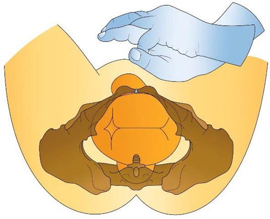
Figure 1. Application of suprapubic pressure.
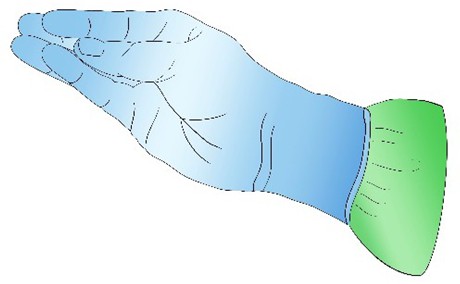
Figure 2. Method of vaginal access.
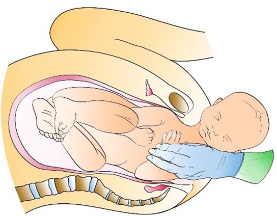
Figure 3. Delivery of the flexed posterior arm.
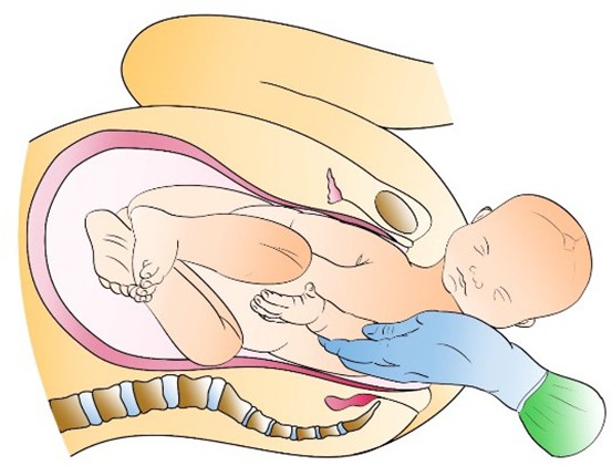
Figure 4. Flexion of the elbow to make the hand accessible.
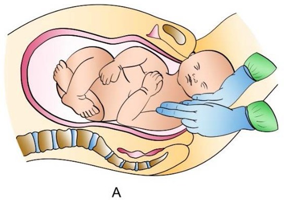
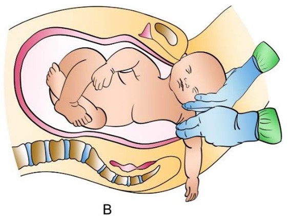
Figure 5. Delivery of the posterior arm by the application of digital traction on the axilla
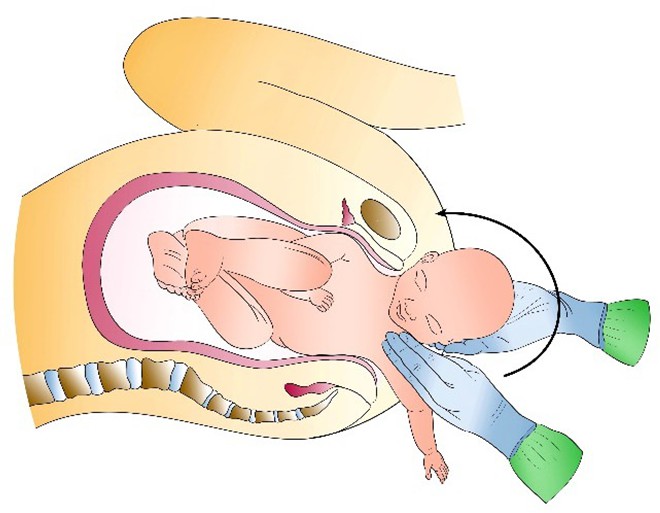
Figure 6. Rotation of the fetus by 180° after delivery of the posterior arm.
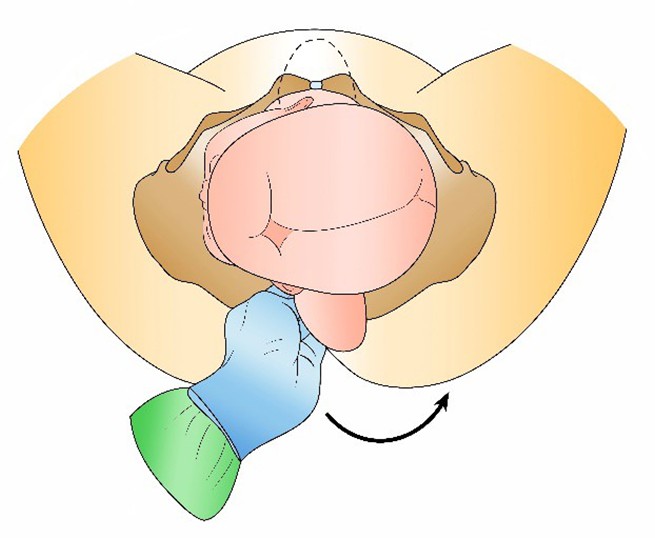
Figure 7. Rotational manoeuvre 1.
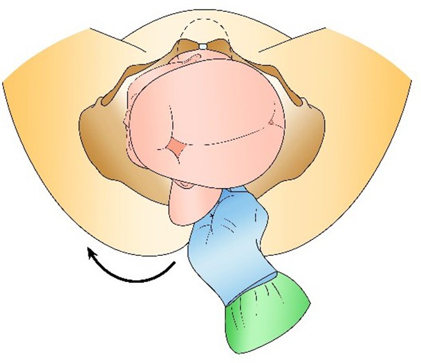
Figure 8. Rotational manoeuvre 2.
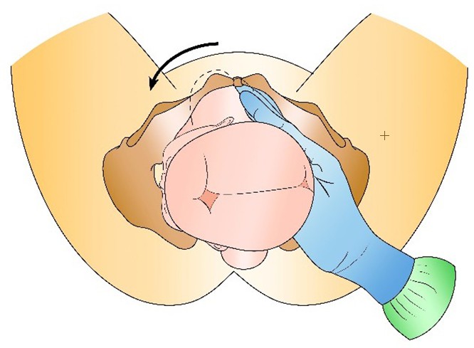
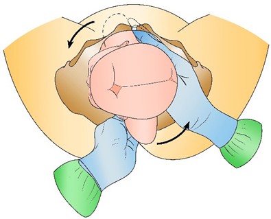
Figure 9. Rotational manoeuvre 3. Figure 10. Rotational manoeuvre 4.
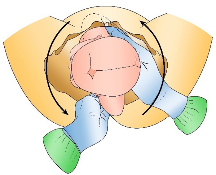
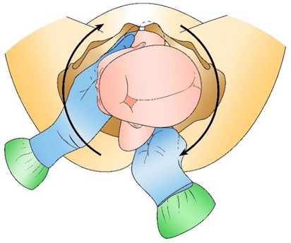
Figure 11. Rotational manoeuvre 5. Figure 12. Rotational manoeuvre 6.