
Recommendations on fetal movements
- All pregnant women should be educated during the antenatal period regarding normal fetal movements. Information on the changing movement patterns as the fetus develops, normal wake/sleep cycles and factors which may modify the mother’s perception of fetal movements should be provided.
Recommendations on fetal movement monitoring
- Clinicians should inform women about the importance of maternal awareness of fetal movements at every antenatal visit in the third trimester.
- Kick-charts are not currently recommended as part of routine antenatal care.
- Women concerned about RFM should be advised to contact their healthcare provider immediately.
Recommendations for the management of a single episode of RFM
- When a woman reports RFM, an assessment of the woman and her fetus should be undertaken as soon as possible, within 2 hours.
- The assessment should include a detailed history of risk factors associated with an increased risk of stillbirth, confirming fetal viability using a handheld doppler and measurement of symphysis-fundal height.
A CTG should be performed to exclude immediate fetal compromise with further evaluation for women with any abnormal CTG pattern.
- Ultrasound assessment of fetal biometry, amniotic fluid volume and umbilical artery dopplers should be considered as part of the preliminary investigation of a woman reporting RFM. Evaluation of fetal morphology should be performed when facilities are available if this has not already been performed.
- Testing for feto-maternal haemorrhage should be considered in the preliminary investigation of women with RFM.
- Biophysical profile (BPP) with non-stressed CTG should be considered in managing high-risk women with RFM.
- In the presence of a normal clinical assessment (including a CTG and ultrasound), if maternal concern of RFM persists, consultant opinion should be sought, and further management should be individualised.
Recommendations for the management of recurrent episodes of RFM
A consultant’s opinion should be sought, and a detailed fetal ultrasound assessment should be performed.
Background
Fetal movement is defined as ‘the maternal sensation of any discrete kick, flutter, swish or roll1. It indicates the normal integrity of the central nervous system and musculoskeletal systems. Quickening is the first sign of fetal movement perceived by a pregnant woman between 18 and 20 weeks of gestation2. There is no universally agreed definition of RFM. However, a significant reduction or alteration in fetal movement, as perceived by the mother, is an important clinical sign of fetal compromise and impending fetal death and has been associated with poor perinatal outcomes3,4. Among women who experienced stillbirth, 55% perceived reduced fetal movement before the diagnosis5. Inappropriate response by clinicians to the maternal perception of RFM is a common contributory factor in stillbirth6.
Normal fetal movements during pregnancy
Most pregnant women feel quickening by 18-20 weeks gestation. Some multiparous women may perceive it as early as 16 weeks of gestation, while some primiparous women may perceive it later than 20 weeks7. The number of spontaneous fetal movements tends to increase until 32 weeks of gestation and plateaus after that until the onset of labour8. By term, the average number of fetal movements per hour is 31, ranging between 16 to 45, with the longest period between movements ranging from 50 to 75 minutes. Fetal movements show diurnal variation, with peaks of activity thought to be in the afternoon and evening periods. During ‘sleep’ cycles, fetal movements are absent.
In a normal, healthy fetus, the ‘sleep’ cycle usually lasts 20-40 minutes and rarely exceeds 90 minutes9.
Factors that influence a woman’s perception of fetal activity
A wide variety of factors influence fetal activity. Women perceive most fetal movements when lying down, fewer when sitting and fewest while standing10. An anterior placenta may decrease the perception of fetal movements before 28 weeks gestation11. Sedating drugs such as alcohol, benzodiazepines, methadone and other opioids cross the placenta and cause a transient reduction in fetal movements12. Cigarette smoking may decrease fetal activity from 30 weeks onwards due to the influence of carbon dioxide in maternal blood on fetal respiration13. Transient reduction in fetal activity and fetal heart rate variability on CTG has been associated with administering corticosteroids for fetal lung maturity over the 2 days following administration14. Fetal malformations such as abnormalities of the central nervous system, muscular dysfunction or skeletal abnormalities are more likely to be associated with reduced fetal movements15. The fetal spine positioned anteriorly may be associated with reduced perception of fetal activity16.
Fetal movement monitoring
Fetal movements should be assessed by maternal perception. The greatest number of fetal movements are perceived when the mother is lying down and in the evening10. Women should be advised to be aware of their baby’s pattern of movements. Current evidence does not support using kick-charts as part of routine antenatal care17. Instructing women to monitor fetal movements may cause increased maternal anxiety18. However, kick-charts are helpful for high-risk pregnancies, especially where there is evidence of FGR19.
Women who are unsure regarding RFM after 28 weeks should be advised to lie on their left side and focus on fetal movements for 2 hours. If they do not feel 10 or more discrete movements within 2 hours, they should immediately contact their healthcare provider. This includes their Family Health Midwife, Medical Officer of Health, Primary Care Unit, Emergency Treatment Unit, Hospital Maternity Ward or Obstetrician. They should be advised not to delay seeking care or wait until the next day for an assessment.
Management of a single episode of RFM
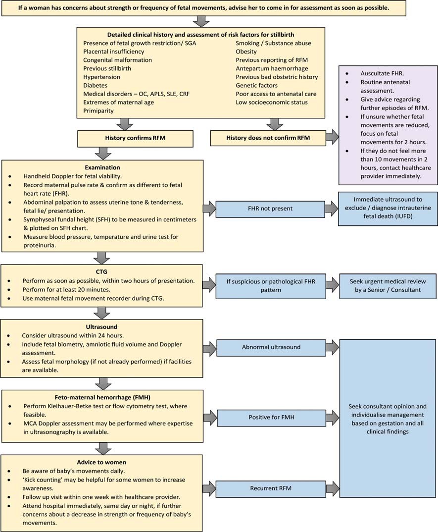
When a woman reports RFM, an assessment of the woman and her fetus should be undertaken as soon as possible, within 2 hours20.
History
A detailed history should be taken, including the duration of RFM and risk factors identification. Factors associated with an increased risk of stillbirth include fetal growth restriction (FGR), small for gestational age fetus (SGA), placental insufficiency, congenital malformation, previous stillbirth, hypertension, diabetes, medical disorders such as obstetric cholestasis (OC), antiphospholipid anti-body syndrome (APLS), systemic lupus erythe-matosus (SLE), chronic renal failure (CRF), extremes of maternal age, primiparity, smoking, substance abuse, obesity, previous reporting of RFM, antepartum haemorrhage, previous bad obstetric history, genetic factors, poor access to antenatal care and low socioeconomic status21.
Examination
Fetal viability must be confirmed using a handheld doppler. If a fetal heartbeat is not detected by hand-held doppler, an ultrasound scan assessment of fetal cardiac activity must be undertaken immediately. The maternal abdomen should be palpated to assess uterine tone, tenderness, and fetal lie/presentation. Assessment of fetal size should be undertaken using symphysis-fundal height (SFH) measurement to detect an SGA fetus. Measurement of blood pressure and a urine test for proteinuria to detect pre-eclampsia is advisable.
Cardiotocography (CTG)
In a woman with a history confirming RFM and over 28 weeks gestation, a CTG should be performed as soon as possible, within 2 hours of presentation, to exclude immediate fetal com-promise22. CTG monitoring of the fetal heart rate for at least 20 minutes provides a means of detecting fetal compromise. For women with any abnormal CTG pattern, immediate assessment by a senior doctor is recommended.
Ultrasound scan
An ultrasound scan should be performed as part of the preliminary investigations in a woman presenting with RFM, particularly for those with persistent RFM despite a normal CTG, risk factors for FGR/ stillbirth, with a clinical suspicion of SGA/ FGR and when the measurement of SFH is likely to be less accurate, such as in those with high body mass index or uterine fibroids.
Ultrasound scan assessment should be arranged preferably within 24 hours and should include evaluation of fetal biometry, amniotic fluid volume and umbilical artery dopplers. A fetal anomaly scan should be arranged if it has not been performed previously if facilities are available20.
Testing for feto-maternal haemorrhage
Testing for feto-maternal haemorrhage (FMH) using the Kleihauer test or flow cytometry test should be considered in the preliminary investigation of women with RFM23. In some women, RFM may be the only history suggesting the possibility of a massive feto-maternal haemorrhage. MCA doppler assessment may be performed where expertise in ultrasonography is available24.
Biophysical profile (BPP)
Although evidence to support the use of biophysical profile (BPP) with non-stressed CTG is limited, it should be considered in the management of high-risk women with RFM25.
Women can be reassured that 70% of pregnancies with a single episode of RFM are uncomplicated26. However, in the presence of a normal clinical assessment (including a CTG and ultrasound), if maternal concern of RFM persists, consultant opinion should be sought, and further management should be individualised.
Management of recurrent episodes of RFM
A consultant should assess women presenting with recurrent episodes of RFM. An ultrasound scan should be undertaken as part of the investigations. The risk of poor perinatal outcomes is higher among these women27. A consultant-led decision to induce labour at term despite normal fetal growth, liquor volume, and CTG should be considered after counselling.
- Optimal management of RFM before 24+0 weeks of gestation
Fetal viability should be confirmed by auscultation by handheld doppler. If the fetal movement has never been felt by 24 weeks of gestation, an ultrasound assessment should be performed to look for evidence of fetal neuromuscular conditions2
- Optimal management of RFM between 24+0 and 28+0 weeks of gestation
Fetal viability should be confirmed by handheld doppler. A comprehensive history should be under-taken to assess for risk factors of stillbirth. Evidence does not support the routine use of CTG. However, an ultrasound scan is recommended as placental insufficiency may present at this gestation.
Algorithm for women presenting with Reduced Fetal Movements (RFM) from 28 weeks gestation
References
- Neldam S. Fetal movements as an indicator of fetal well-being. Dan Med Bull. 1983; 30(4): 274-8.
- Rayburn WF. Fetal body movement monitoring. Obstet Gynecol Clin North Am. 1990; 17(1): 95-110.
- Grant A, Elbourne D, Valentin L, Alexander S. Routine formal fetal movement counting and risk of antepartum late death in normally formed singletons. Lancet. 1989; 2(8659): 345-9.
- Harrington K, Thompson O, Jordan L, Page J, Carpenter RG, Campbell S. Obstetric outcome in women who present with a reduction in fetal movements in the third trimester of pregnancy. J Perinat Med. 1998; 26(2): 77-82.
- Efkarpidis S, Alexopoulos E, Kean L, Liu D, Fay T. Case-control study of factors associated with intrauterine fetal deaths. Med Gen Med. 2004; 6(2): 53.
- Saastad E, Vangen S, Froen JF. Suboptimal care in stillbirths – a retrospective audit study. Acta Obstet Gynecol Scand. 2007; 86(4): 444-50.
- Marsal K. Ultrasonic assessment of fetal activity. Clin Obstet Gynaecol. 1983; 10(3): 541-63.
- Natale R, Nasello-Paterson C, Turliuk R. Longitudinal measurements of fetal breathing, body movements, heart rate, and heart rate accelerations and decelerations at 24 to 32 weeks of gestation. Am J Obstet Gynecol. 1985; 151(2): 256-63.
- Patrick J, Campbell K, Carmichael L, Natale R, Richardson B. Patterns of gross fetal body movements over 24-hour observation intervals during the last 10 weeks of pregnancy. Am J Obstet Gynecol. 1982; 142(4): 363-71.
- Minors DS, Waterhouse JM. The effect of maternal posture, meals and time of day on fetal movements. Br J Obstet Gynaecol. 1979; 86(9): 717-23.
- Neldam S, Jessen P. Fetal movements registered by the pregnant woman correlated to retrospective estimations of fetal movements from cardioto-cographic tracings. Am J Obstet Gynecol. 1980; 136(8): 1051-4.
- Castillo RA, Devoe LD, Ruedrich DA, Gardner P. The effects of acute alcohol intoxication on biophysical activities: a case report. Am J Obstet Gynecol. 1989; 160(3): 692-3.
- Richardson BS, O’Grady JP, Olsen GD. Fetal breathing movements and the response to carbon dioxide in patients on methadone maintenance. Am J Obstet Gynecol. 1984; 150(4): 400-5.
- Magee LA, Dawes GS, Moulden M, Redman CW. A randomised controlled comparison of beta-methasone with dexamethasone: effects on the antenatal fetal heart rate. Br J Obstet Gynaecol. 1997; 104(11): 1233-8.
- Lin CC, Adamczyk CJ, Sheikh Z, Mittendorf R. Fetal congenital malformations. Biophysical profile evaluation. J Reprod Med. 1998; 43(6): 521-7.
- Fisher M. Reduced fetal movements: a research-based project. The British Journal of Midwifery. 1999; 7: 733-7.
- Mangesi L, Hofmeyr GJ, Smith V, Smyth RM. Fetal movement counting for assessment of fetal wellbeing. Cochrane Database Syst Rev. 2015; 2015(10): Cd004909.
- Draper J, Field S, Thomas H, Hare MJ. Womens’ views on keeping fetal movement charts. Br J Obstet Gynaecol. 1986; 93(4): 334-8.
- Haws RA, Yakoob MY, Soomro T, Menezes EV, Darmstadt GL, Bhutta ZA. Reducing stillbirths: screening and monitoring during pregnancy and labour. BMC Pregnancy Childbirth. 2009; 9 Suppl 1(Suppl 1): S5.
- Tveit JVH, Saastad E, Stray-Pedersen B, Bordahl PE, Flenady V, Fretts R, et al. Reduction of late stillbirth with the introduction of fetal movement information and guidelines – a clinical quality improvement.
- Confidential Enquiry into Stillbirths and Deaths in Infancy(CESDI). 8th Annual Report. London: Maternal and Child Health Research Consortium; 2001.
- Ahn MO, Phelan JP, Smith CV, Jacobs N, Rutherford SE. Antepartum fetal surveillance in the patient with decreased fetal movement. Am J Obstet Gynecol. 1987; 157(4 Pt 1): 860-4.
- Wylie BJ, D’Alton ME. Fetomaternal hemorrhage. Obstet Gynecol. 2010; 115(5): 1039-51.
- Eichbaum M, Gast AS, Sohn C. Doppler sonography of the fetal middle cerebral artery in the management of massive fetomaternal hemorrhage. Fetal Diagn Ther. 2006; 21(4): 334-8.
- Dayal AK, Manning FA, Berck DJ, Mussalli GM, Avila C, Harman CR, et al. Fetal death after normal biophysical profile score: An eighteen-year expe-rience. Am J Obstet Gynecol. 1999; 181 (5 Pt 1):
1231-6.
- Heazell AE, Sumathi GM, Bhatti NR. What investigation is appropriate following maternalperception of reduced fetal movements? J Obstet Gynaecol. 2005; 25(7): 648-50.
- O’Sullivan O, Stephen G, Martindale E, Heazell AE. Predicting poor perinatal outcome in women who present with decreased fetal movements. J Obstet Gynaecol. 2009; 29(8): 705-10.
- Rayburn WF, Barr M. Activity patterns in malformed fetuses. Am J Obstet Gynecol. 1982; 142(8): 1045-8.