Introduction
Maternal collapse is a rare but life-threatening event. Whilst maternal collapse is such an uncommon event, the consequences are potentially devastating, therefore it is essential that the clinical team are skilled in initial effective resuscitation techniques. This guideline aims to highlight the evidence on immediate resuscitation following maternal collapse during pregnancy in a hospital.
The Royal College of Obstetricians and Gynaecologists has defined maternal collapse as “an acute event involving the cardio-respiratory systems and/or brain, resulting in a reduced or absent conscious level (and potentially death), at any stage in pregnancy and up to 6 weeks after delivery”.
Outcome following maternal collapse depends on prompt and immediate resuscitation.
- Key recommendations for immediate resuscitation
- An obstetric modified early warning score chart should be used for all women undergoing observation, to allow early recognition of the woman who is becoming critically ill.
- Maternal collapse can result from a number of causes. A systematic approach should be taken to identify the cause and treat accordingly to improve the outcome (See 3).
- It is essential that anyone involved in the resuscitation of pregnant women is aware of the physiological differences (See 4, Figure 2).
- Aortocaval compression significantly reduces cardiac output by up to 30-40% from 20 weeks of gestation onwards. This also reduces the efficacy of chest com-pressions during resuscitation.
- Pregnant women become hypoxic more readily and make ventilation more difficult.
- Difficult intubation is more likely in pregnancy due to laryngeal oedema, large breasts and weight gain.
- Pregnant women are at an increased risk of aspiration due to the relaxation of the lower esophageal sphincter, raised intra-abdominal pressure and delayed gastric emptying.
- Maternal collapse resuscitation should follow the resuscitation guidelines using the standard ABCDF approach, with some modifications for maternal physiology, in particular relief of aortocaval compression.
- If the airway is clear and there is no breathing, chest compressions should be commenced immediately (See 4).
- Early intubation is essential by the most experienced clinician present.
- A left lateral tilt of the woman from head to toe at an angle of 15-30o on a firm surface will relieve aortocaval compression (See appendix: Figure 3).
2.4.4. Manual displacement of the uterus to the left is effective in relieving aortocaval compression in women above 20 weeks’ gestation. (See appendix: Figure 3, 4).
- Two wide-bore cannulae (minimum 16 gauge) should be inserted as soon as possible. Intravenous access should be achieved at or above the level of the diaphragm.
- Perimortem caesarean section (PMCS) should be performed by the most experienced clinician available, if there is no response to CPR at the 5th minute.
- Perimortem caesarean section should be performed where resuscitation is taking place, without moving the woman.
- The operator should use the incision that will facilitate the most rapid access. In terms of the best incision to use, a midline abdominal incision and a classic uterine incision will give the most rapid access. (See appendix: Figure 5).
- With the ‘best interests of the patient’ this procedure would be carried out without consent.
- No anaesthetic or analgesic is required to perform a PMCS.
2.7. The same defibrillation energy levels should be used for shockable rhythm as in a non-pregnant woman. (4J/kg BW)
2.8 Early involvement of an appropriately skilled anesthetist to attend the immediate resuscitation remains the best clinical practice.
Causes of maternal collapse
- Major obstetric haemorrhage: include postpartum haemorrhage, major antepartum haemorrhage from placenta praevia, placental abruption, uterine rupture and ectopic pregnancy. Also concealed haemorrhage due to cesarean section, splenic artery rupture or hepatic rupture.
- Thromboembolism
- Amniotic fluid embolism (AFE)
- Cardiac disease leading to cardiac arrest
- Sepsis
- Drug toxicity
- Eclampsia
- Intracranial haemorrhage
- Anaphylaxis
- 4H, 4T (See appendix; Table 1)
The physiological and anatomical changes in pregnancy that affect resuscitation
- Aortocaval compression significantly reduces cardiac output from 20 weeks of gestation onwards and the efficacy of chest compressions during resuscitation. As a consequence, cardiac output is reduced by up to 30-40% (See appendix: Figure 2).
- Changes in lung function, diaphragmatic splinting and increased oxygen consumption make pregnant women become hypoxic more readily and make ventilation more difficult.
- Difficult intubation is more likely in pregnancy due to laryngeal oedema, large breasts and weight gain.
- Pregnant women are at an increased risk of aspiration due to the relaxation of the lower esophageal sphincter, raised intra-abdominal pressure and delayed gastric emptying.
Cardio-vascular changes in pregnancy include:
- Increased cardiac output and hyperdynamic circulation of pregnancy mean that large volumes of blood can be lost rapidly, especially from the uterus which receives 10% of the cardiac output at term.
- Aortocaval compression reduces preload and significantly impede the efficacy of chest compression.
- Common, reversible causes of maternal cardio-pulmonary arrest should be considered throughout the resuscitation process. (See appendix Table 1, Figure 1)
- Resuscitation efforts should be continued until a decision is taken by the consultant obstetrician and consultant anesthetist to discontinue resuscitation efforts.
Optimal initial management of maternal collapse
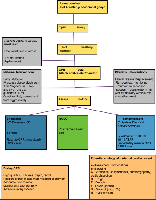
- Alert the cardiac arrest team.
- Effective chest compression in a collapsed pregnant woman is different from a non-pregnant woman.
- A left lateral tilt of the woman from head to toe at an angle of 15-30° on a firm surface will relieve aortocaval compression in the majority of pregnant women and still allow effective chest compressions to be performed in the event of cardiac arrest (See appendix: Figure 3).
- Manual displacement of the uterus to the left is effective in relieving aortocaval compression in women above 20 weeks’ gestation or where the uterus is palpable at or above the level of the umbilicus. This permits effective chest compressions in the supine position in the event of cardiac arrest (See appendix: Figure 3, 4).
- In cases of major trauma, the spine should be protected with a spinal board before any tilt is applied. Thereafter, manual displacement of the uterus should be used in this event.
- Intubation in an unconscious woman with a cuffed endotracheal tube should be performed immediately by an experienced medical officer.
- Early intubation is essential by the most experienced clinician present.
- Bag and mask ventilation or insertion of a simple supraglottic airway should be undertaken until intubation can be achieved.
- Supplemental high flow oxygen via nasal cannula should be administered as soon as possible to counteract rapid deoxygenation. This should be maintained until intubation.
- Two wide-bore cannulae (minimum 16 gauge) should be inserted as soon as possible. If peripheral venous access is not possible, early consideration of central venous access or venous cutdown should be considered.
- Intravenous access should be achieved at or above the level of the diaphragm.
- There should be an aggressive approach to volume replacement, although caution should be exercised in the context of pre-eclampsia or eclampsia.
- Initial recommended volume replacement is 1 liter over 20 minutes.
- The same defibrillation energy levels should be used for shockable rhythm as in a non-pregnant woman (4J/kg BW). Correct positioning of the pads is essential for successful defibrillation to take place (See appendix: Figure 6).
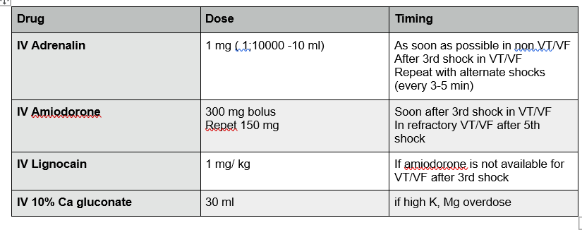
- There should be no alteration in algorithm drugs or doses used in the Resuscitation protocols (See appendix: flow chart 1, Table 3).
Perimortem caesarean section (PMCS)
- In women over 20 weeks of gestation, if there is no response to correctly performed CPR within 4 minutes of maternal collapse or if resuscitation is continued beyond this, then PMCS should be undertaken to assist maternal resuscitation. Ideally, this should be achieved within 5 minutes of the collapse.
PMCS should not be delayed by moving the woman. It should be performed where maternal collapse has occurred and resuscitation is taking place.
- The operator should use the incision, which will facilitate the most rapid access. This may be a midline vertical incision or a suprapubic transverse incision. In terms of the best incision to use, a midline abdominal incision and a classic uterine incision will give the most rapid access to deliver the fetus.
- A fixed blade scalpel and umbilical cord clamps (or alternative ligatures) should be available on the resuscitation trolley in all areas where maternal collapse may occur, including the accident and emergency department.
- With no circulation, blood loss is minimal, and no anaesthetic is required to perform a PMCS.
- If resuscitation is successful following birth, there should be prompt transfer to an appropriate environment at that point, as well as anaesthesia and sedation, to control ensuing haemorrhage and complete the operation. With the ‘best interests of the patient’ this procedure would be carried out without consent.
- Research groups found that 69% of infants survived when PMCS was performed within 5 minutes. Delivery of the fetus and placenta reduces oxygen consumption, improves venous return and cardiac output, facilitates chest compressions and makes ventilation easier.
- Outcomes for mothers and babies depend on the cause of collapse, gestational age and access to emergency care, with survival rates being poorer if the collapse occurs out of hospital. In maternal cardiac arrest maternal survival rates of over 50% have been reported in UK.
Training
- All generic life support training should consider the adaptation of CPR in pregnant women. All maternity staff should have annual formal multi-disciplinary training in generic life support and the management of maternal collapse.
- Life support training improves resuscitation skills. Small group multidisciplinary interactive practical training is recommended to improve the management of maternal collapse.
Clinical governance
- Documentation – Accurate documentation in all cases of maternal collapse, whether or not resuscitation is successful, is essential.
- Debriefing is recommended for the woman, her family and the staff involved in the event.
- Incident reporting.
Essentials for emergency obstetrics care should be provided to all base hospitals and above
- In order to reduce maternal mortality, comprehensive Emergency Obstetric Care (EmOC) must be available and accessible to all women.
- The components of basic and comprehensive EmOC as recommended by World Health Organization (See appendix: Table 2).
Appendix
Table 1
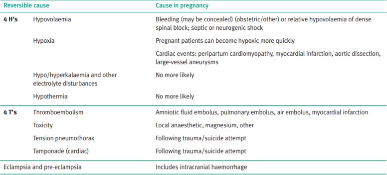
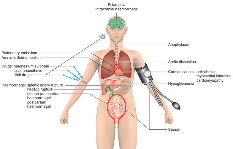
Figure 1. Causes of maternal collapse.
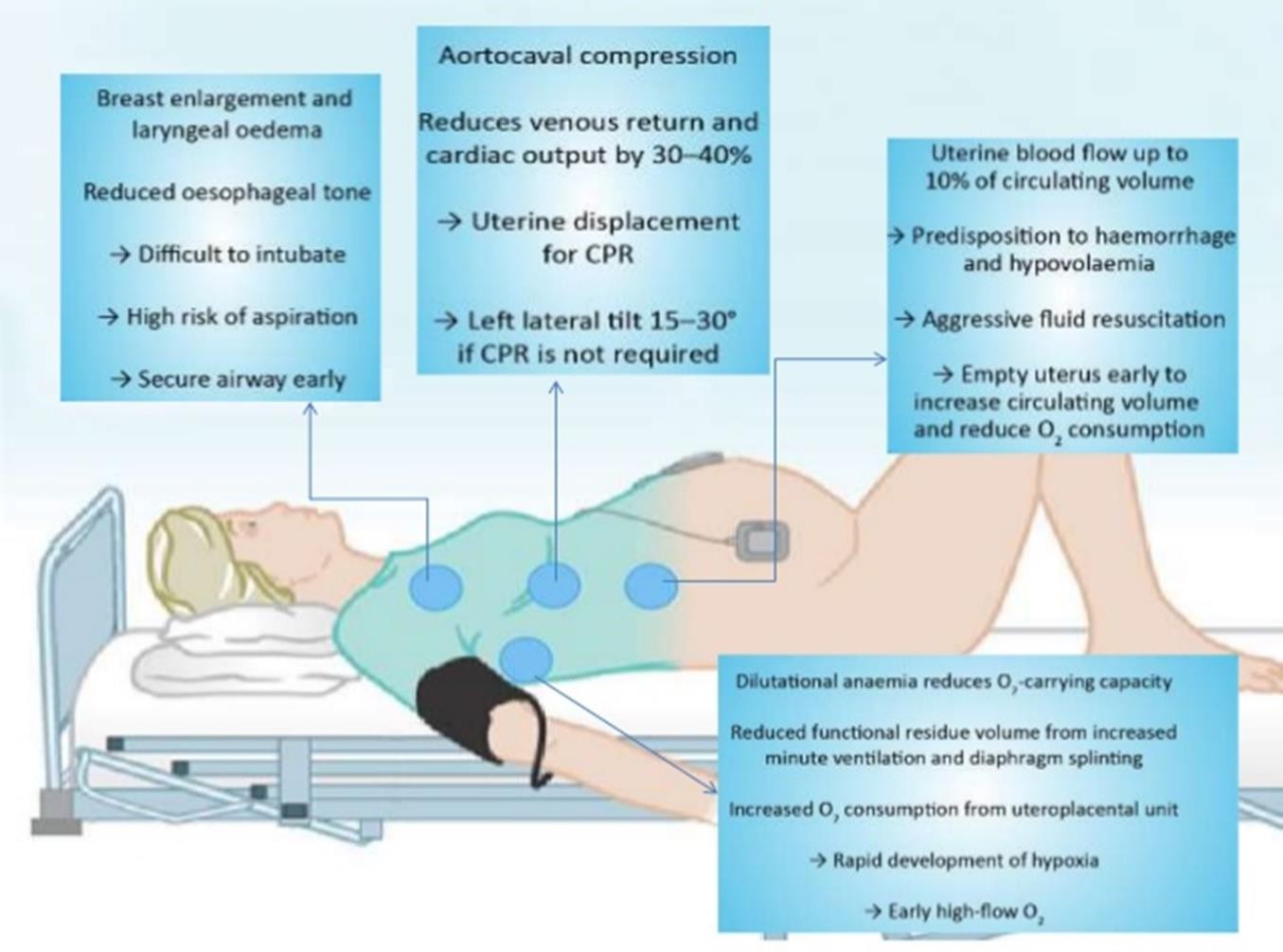
Figure 2. Physiological changes in pregnancy and the effects on resuscitation.
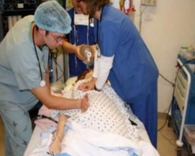

Figure 3. Relieving the aorto-caval compression.
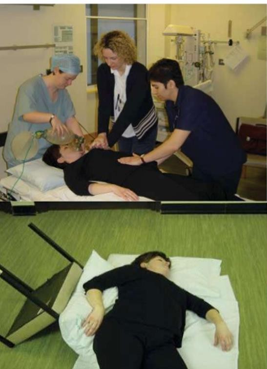
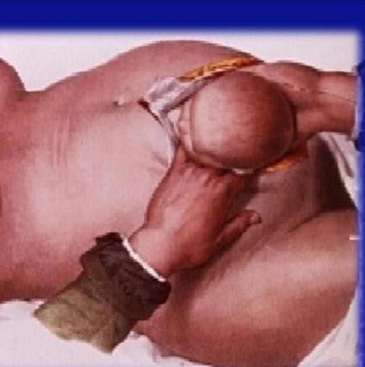
Figure 4. Relieving aorto-caval compression Figure 5. Perimortem cesarean section. by uterine displacement and 30º wedge.
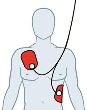
Figure 6. Position of defibrillator pads: Right under the clavicle, Left under the at 5th intercostal space mid axillary line.
Table 2. Basic and comprehensive EmOC
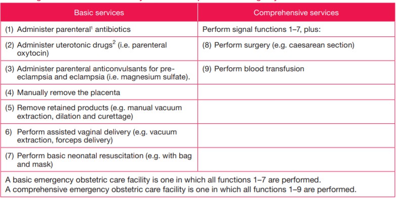
Table 3. Common drugs used during maternal cardiac arrest
Flow chart
Drugs used during maternal cardiac arrest