
Introduction
Cervical cancer is the leading gynaecological malignancy in Sri Lanka. Among women, it is the second most common cancer, where the first is breast cancer. There were 1136 new cases reported in 2018. In that year, 643 cervical cancer deaths were reported. This shows most of the cancers have a late-stage presentation and nearly half of them die despite treatment. Similar numbers have been observed in each year and it is ranked as the second leading cause of female cancer deaths in Sri Lanka also1.
Cervical cancer is unique compared to many other cancers. It has a well-defined pre-invasive state, which could be detected through cervical screening. Countries with well-established screening programmes have reduced cancer incidence significantly over the past decades. For example in the United Kingdom (UK), cancer incidence has reduced by 75% since the introduction of the successful national screening programme in the 1970s2.
Cervical screening will also help in the early detection of cancer. Most of the cancers detected through the cervical screening programme are of early stages, which can be successfully treated. On the contrary,
large numbers of the cancers presented in Sri Lanka are of late stages which have less success of cure, despite intense treatment options like radical hysterectomy and radiotherapy, etc. National cancer hospital of Maharagama is one of the centers providing advanced surgical options for these patients, and around 100 patients will have Wertheim's hysterectomy for cervical cancer annually in this hospital alone.
Conventional cytology-based cervical smears have low to moderate sensitivity (53% to detect Cervical Intraepithelial Neoplasia (CIN) 2+) and specificity around 96% in detecting abnormal cells3. Smears can be done in the out-patient setting simply using a Cusco’s bivalve speculum and Ayer’s Spatula without the need for anesthesia or sedation. Processing of the smears needs laboratory facilities and trained personnel is needed to interpret them. This is a significant challenge and limitation of our smear programme.
At present, our smear programme is coordinated by the Well Women Clinic – Family Health Bureau centrally and the Public Health Nursing Sisters peripherally under the guidance of the Medical Officer of Health (MOH) in that particular region. The pap smear programme has an uptake of 30-50% of the target population in different districts.
a Accredited Colposcopist and Trainer BSCCP-UK, Consultant Gynaecologist, Hywel Dda University Health Board, West Wales, UK
b Consultant Gynaecological Oncologist, National Hospital, Kandy, Sri Lanka
c Consultant Gynaecological Oncologist, National Cancer Institute (Apeksha Hospital), Maharagama, Sri Lanka
d Consultant Gynaecological Oncologist, Teaching Hospital, Anuradhapura, Sri Lanka
e Sub-speciality Trainee in Gyanaecological Oncology, National Cancer Institute (Apeksha Hospital), Maharagama, Sri Lanka
The World Health Organisation (WHO) recommends screening for cervical cancer to be started at the age of 30 years and to continue until the age of 65 years. Currently in Sri Lanka screening is done targeting two age cohorts; 35 and 45 years4.
However, considering the rising incidence of cervical cancers, women who had been sexually active can be offered cervical smear screening 3 to 5 years since their first sexual intercourse.
In the UK, the first smear is done at the age of 25 and if Human Papilloma Virus (HPV) is negative, threeyearly follow-up smears are done until the age of 50. After 50 years, the interval becomes 5 years and the women will be discharged from the smear programme at the age of 64 maximally. If the last 2 smears are HPV negative, she will be ceased from the cervical screening programme.
Classification of smears
Bethesda system is currently used to define cervical cytology smears. The following categories are included in the modified classification adapted for Sri Lanka4.
Cytology based classification
1. Negative for Intraepithelial Lesion or Malignancy (NILM)
2. Low grade Squamous Intraepithelial Lesion (LSIL)
3. High grade Squamous Intraepithelial Lesion (HSIL)
4. Atypical Squamous Cells of Undetermined Significance (ASCUS) which is also known as borderline smear. This is further subdivided into two categories
- Atypical Squamous Cells of Undetermined Significance – Low grade (ASCUS – Low grade)
- Atypical Squamous Cells of Undetermined Significance – High grade (ASCUS – High grade)
5. Glandular cell atypia
6. Benign endometrial cells in a woman > 40 years (incidental finding in smears sometimes)
7. Suspicious of invasive cancer either squamous or glandular
8. Inadequate smears (cells are not adequate enough for interpretation)
More recently high risk (HR) HPV virus DNA detection was introduced to the screening. This test does not use cytology as the primary marker and instead use a cohort of HR HPV DNA probes to detect their presence. The result is simply either positive or negative for high risk HPV.
Most of the developed countries are rapidly moving towards primary HPV screening replacing the conventional cytology-based screening. In Sri Lanka, HR HPV screening test is available in the private sector and whenever possible its use has been discussed in this guidance. However, our primary screening is still cytology and hence its use is mostly discussed.
Cervical cancer screening methods
1. HPV DNA based screening
2. Cervical cytology based screening
HPV DNA based screening
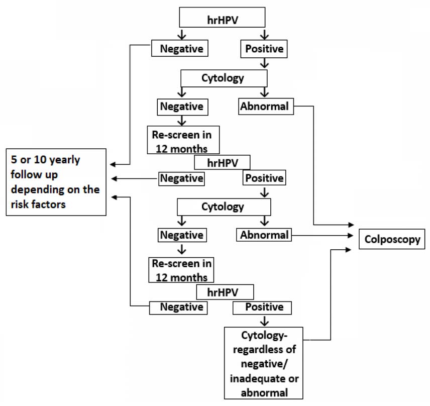
- If the HR-HPV DNA is negative, the patient can be referred for the routine review in 5 years in the presence of risk factors (Annexure 1).
- All patients with positive HPV DNA should have cytology tested.
- Positive HR-HPV DNA and abnormal cervical cytology warrants colposcopic examination.
- In cases where the cervical smear is negative in positive HR-HPV DNA patients, HR-HPV DNA status should be rechecked in 12 months after the first HR-HPV DNA testing.
If the cytology and 12 months repeat HR- HPV DNA both are negative, patients should be screened after 10 years or if they are above 45 years of age further screening is not necessary.
- Colposcopy is indicated in
1st positive HR-HPV DNA with abnormal cytology
Positive HR HPV DNA tests despite the negative cytology for 2 consecutive years
- If the colposcopy is negative, they are followed up with three-yearly cervical smears. If HR-HPV becomes negative patient can be discharged for routine smears. If cytology becomes positive follow the appropriate pathways.
Cervical cytology smear based screening
- The current recommendation is for women to have at least two smears done between the age of 35 to 45 years. This is based on a WHO document that gives guidance to cervical screening in low and middle-income countries.
- However if the facilities are available, the screening can be arranged more frequently (3-5 years) in high-risk populations (Annexure 1) or geographical areas with the high incidence and especially in the areas of high prevalence of HIV.
- If women wish the more regular screening we recommend this to be done ideally 3 yearly until the age of 50 years and 5 yearly afterward up to the age of 64 years.
- However, screening intervals can be modified according to the individual circumstances and the availability of facilities.
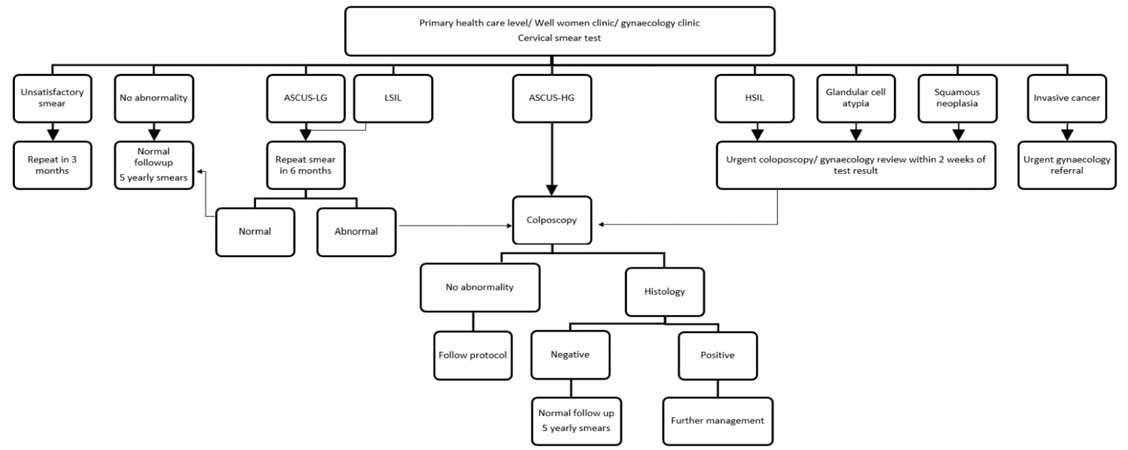
- When the smear is inadequate it should be repeated after 3 months.
- If the smear is ASCUS-LG (Borderline LG) or LSIL (mild dyskaryosis) cervical cytology smear test needs to be repeated in 6 months’ time.
- If the repeat cervical smear in 6 months is negative, the patient will be referred for routine follow-up.
- If the repeat cervical cytology smear is abnormal after 6 months, ideally the patient should be referred for colposcopy. Colposcopic assessment and necessary biopsies will decide further management. Follow-up care should be planned appropriately.
- However, if colposcopy referral is not feasible, we would suggest continue smears in 6 monthly intervals. It is difficult to support this with evidence. However, considering the practical difficulties, especially in low resource areas, this would be an option to consider.
- If the abnormality resolves, the patient can be reverted back to standard screening and if she develops high grade disease (HSIL) urgent referral to a unit where colposcopy facilities are available is recommended.
- After two years of follow-up, if the low grade smear abnormality is persistent, either referral to a colposcopy clinic or treatment at the local center should be considered.
- Immediate colposcopy is indicated if the smear is
ASCUS-HG
HSIL
Glandular abnormaity
Suspicious of invasive cancer
Positive predictive value for high grade lesions is high in colposcopy (60-80%). However, for low-grade disease this is around 15%. Colposcopy proforma should be used for all the patients (the following data should be included for every patient – Annexure 2). Directed punch biopsies or excisional biopsies should be done when indicated and follow-up care should be planned appropriately.
If colposcopy does not find any abnormality, it becomes a negative colposcopy. It is important to visualize the entire transformation zone before confirming colposcopy as negative. The abnormality can be within the endocervical canal especially in menopausal women. Sometimes VAIN (Vaginal intraepithelial neoplasia) can give rise to HSIL smears, hence examination of the vagina with iodine is important if the cervix appears normal.
If the colposcopy is negative in a HG smear, ideally the case has to be discussed in a Colposcopy MDT meeting. All the smears and histology are reviewed by a panel of experts.
If the panel decision is the same, diagnostic LLETZ will be the next option.
If LLETZ is not available, diagnostic cone biopsy could be offered if the woman has completed her family.
If the cervix appears abnormal or suspicious of invasive cancer, urgent colposcopy should be considered if possible, irrespective of the smear history. At least an urgent gynecology referral should be done. Clinically suspicious symptoms of cervical cancer like post-coital bleeding, persistent discharge despite treatment should more appropriately be referred to general gynaecologists.
The psychological impact of screening
Abnormal smears and colposcopy visits generate a high degree of anxiety and fear among women and they should be treated with respect and dignity, addressing
their specific concerns all the time. Some believe finding abnormal cells in smear or histology is equivalent to cancer. Patients should be explained in detail the difference between finding pre-cancer cells and cancer cells. Reassurance is very important with clear management plans.
Some patients have anxieties about harboring HPV virus and this could be considered as a sexually transmitted infection. This could even lead to relationship difficulties, and one has to be sensitive when dealing with these issues. Patients should be reassured and educated about HPV as a community-acquired virus. It does not cause symptoms of typical STD like chlamydia or syphilis. Most of the time it is innocuous but at times it will cause damage to the cells of the cervix, which could lead to cancer and, it should be looked into in more detail.
An excisional form of biopsy (LLETZ or cone biopsy) is recommended
1. When most of the ectocervix is replaced with high-grade abnormality.
2. When low-grade colposcopic change is associated with high-grade dyskaryosis (HSIL) severe or worse.
3. When a lesion extends into the canal – sufficient canal must be removed with endocervical extension of the abnormality.
Indications for cold-knife conization5
1. Glandular lesions suspected on cytology and/ or detected on histology
2. Evidence of microinvasive carcinoma
3. High-grade squamous lesions (HSIL) in which the upper limit of the lesion is not visible
4. Disparity between diagnostic modalities
5. Treating high-grade lesions not suitable for ablation in settings where LLETZ facilities are not available
In a selected group of patients locally destructive ablative treatment is indicated. In Sri Lanka cryo cautery is the only option used at present and if used it should only be used for the low grade disease (CIN 1). Generally, CIN 1 can be managed conservatively as the spontaneous resolution of this type of lesion is high. The risk of progression to cancer is also very low. If
treatment is needed for persistent disease “Doublefreeze-thaw-freeze technique” must be used. This method is not suitable to treat high-grade diseases.
Cold coagulation or thermal coagulation is another ablative method used to treat CIN in colposcopy. This method is not available in Sri Lanka and it has been widely used in many other developed and middleincome countries. Heated probe (1200C for 30 seconds) is used to destroy tissue and usually is done under local anesthesia.
Pre-requisites for ablative treatments
1. Entire transformation zone should be visualized
2. No evidence of glandular abnormality
3. No evidence of invasive disease
4. No evidence of glandular crypt involvement
5. No major discrepancy between cytology and histology
6. No history of post-coital bleeding
7. No previous ablative or excisional treatment
Excisional methods like LLETZ and cone biopsy are associated with the risk of preterm labour due to cervical insufficiency. The risk is around 2-3% after a single LLETZ procedure. After two LLETZ procedures, this rises up to 20%. Obviously, cone biopsies cause more damage to the cervix and the risk could be much higher. If the woman wishes to preserve fertility, ablative method could be considered. For high-grade disease, only cold coagulation is the choice but unfortunately, this technique is not yet available in Sri Lanka.
All women must have had a histological diagnosis established by punch biopsies from cervix before destructive therapy, eg. Cryo cautery or Cold Coagulation.
CIN follow-up after treatment
Follow-up can be arranged based on the availability of smears/ high risk HPV DNA test.
HR HPV DNA based follow up
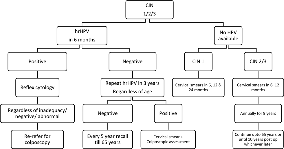
- If HR HPV testing is available it should be done in 6 months in all CIN 1, 2 and 3 patients after the treatment. This test is called ‘Test of cure’ (TOC).
- If 6 monthly smear is negative for HR HPV, the patient can be discharged for routine smears. If HR HPV is still positive she will need colposcopy to assess her cervix. If colposcopy shows no evidence of residual CIN, she can be discharged for routine smears.
- The advantage of HR HPV test is, if negative, repeat test can be done in 3 years regardless of the age. If a test done after 3 years is negative, the patient can be reviewed every 5 years until 65 years.
- If patient becomes positive for HR HPV in 3 years following the guidance in the flowchart.
Cytology based follow up
- In treated cases, if cytology-based screening is followed, for CIN 1 and 2, cervical cytology smears to be taken in 6, 12 and 24 months following the treatment.
- For CIN 3, two cervical smears should be done at 6 monthly intervals first. The screening has to be continued annually for 9 years. Afterward, patients can be referred for routine 5 yearly recall.
Management of abnormal cervical smear in pregnancy
- If a healthy female is due her for routine recall during pregnancy, it can be deferred until 3 months post-delivery.
- However, if they are due screening for previously abnormal smears follow up but defaulted or missed it, cervical smear with or without colposcopy can be considered.
- If an abnormal smear needs colposcopy and the woman is found to be pregnant, this can be performed. This is especially the case if the smear is high grade, Glandular, or suspected invasive disease.
- Colposcopy in the late first trimester or early second trimester should be offered in cases with high-grade smears during the pregnancy.
- Patients with low-grade smears can wait until the patient completes their pregnancy.
- Biopsy during pregnancy is only indicated in clinically or colposcopically suspected invasive
disease. A diagnostic punch biopsy is not recommended as it is not reliable to exclude invasion.
- Otherwise, testing with acetic acid and iodine will alone suffice.
- Good record keeping and pictures should be included in the notes if possible.
- Directed biopsies and treatment of CIN are usually deferred until the post-partum period.
- Those who had colposcopy in pregnancy for an abnormal smear or biopsy-proven CIN, have to undergo repeat colposcopic assessment at 3 months post-partum.
Management of Cervical Glandular Intraepithelial Neoplasia (CGIN)
- CGIN should undergo colposcopy to rule out co-existing squamous abnormalities.
- Endometrial biopsy should be done in high grade glandular neoplasia.
- Excision is the treatment of choice. Ablative treatments are contraindicated in glandular abnormalities.
- Punch biopsies are not recommended and excisional biopsy (e.g. LLETZ) including the endocervical canal can reliably diagnose the lesion.
- Factors including the age, type of transformation zone and fertility wishes have to be considered for the depth of excision.
- In women of the reproductive age group who wish to be pregnant in the future, LLETZ is the treatment of choice and the targeted depth of the loop is kept up to 10 mm. If the woman has completed family the depth of the loop will be around 25mm. Cone biopsy is an option for these women if colposcopy facilities are not available.
- In case of incomplete excision margins, reexcision can be done.
- A hysterectomy is an option in selected cases.
Post hysterectomy follow up
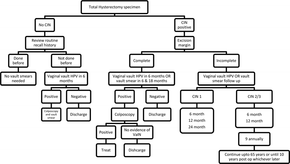
Hysterectomy without CIN
- Smear history should be reviewed in all the patients attending for hysterectomy due to
various reasons. Whenever possible, every attempt should be made to do the smear prior to the hysterectomy if she did not have routine smears.
- Patients who had a normal routine recall and no CIN in their hysterectomy specimen can be discharged from follow-up.
- Those who had no routine recall, and had no smears done prior to hysterectomy, should undergo vaginal vault HPV in 6 months. Patients having negative vault HPV can be discharged. However, those who are having positive vaginal vault HPV/cytology should undergo colposcopy.
- During colposcopy, the vagina should be examined for VAIN with iodine testing.
Hysterectomy specimen with CIN
- In cases with complete excision margins, vaginal vault HPV should be done in 6 months. Those who had negative HPV can be discharged from the follow-up. In cases of positive vaginal vault HPV, colposcopy should be done and treat if abnormalities are detected. However, if there is no evidence of vaginal intraepithelial neoplasia (VaIN), they can be discharged from the followup.
- In case of incomplete excision margins seen in the hysterectomy specimen, patients need to be followed up with HPV testing for at least 6 and 18 months. A positive test will warrant a colposcopy referral.
- If no HPV testing is available, please refer to the flow chart-3 “CIN follow up after treatment”.
Contraception and vaccination
- There is no contraindication for hormonal contraception in the presence of abnormal cervical smears.
- Safe sex using condoms have shown to reduce the risk of transmission of HPV among sexually active populations. However, once a woman acquires HPV her own immune system should eliminate it as there is no cure.
- HPV vaccination of young children before they start sexual activity has been successfully implemented in Sri Lanka. HPV vaccination has shown to be highly effective against acquiring HR HPV in sexually active populations.
Clinical governance
- Considering the resources and observed changing patterns of sexual debut, bringing down the first cervical screening at least to the age of 30 years is recommended in contrast to the previous practice of age of 35 years. However, starting cervical smear screening 3 years following the sexual debut on the patient’s demand needs to be accepted.
- Cervical smear screening interval is recommended for every 10 yearly, however 5 year interval is recommended in the presence of risk factors up to the age of 65 years.
- Colposcopy assessment should be done by a clinician trained in colposcopy. There are few centers in Sri Lanka offering colposcopy services and these facilities should be expanded. Training workshops should be conducted on regular basis to train and update the knowledge and skills of doctors.
- Cervical smears showing HSIL, glandular cell atypia and squamous cell neoplasia needs urgent colposcopy within 2 weeks of test result.
- However, when easy access to colposcopy is not possible, proceeding for excisional biopsy procedures could be considered. Hysterectomy could be considered in selective cases when high grade disease either squamous or glandular when family has been completed, after excluding invasive disease.
- Colposcopy during pregnancy should be done by senior Colposcopist with experience.
- Patients who have undergone a subtotal hysterectomy still need to have followed up cervical smears or HPV DNA testing as for the protocol.
- All patients should have a negative cervical smear within the screening interval or preoperatively, before undergoing hysterectomy.
- Diagnostic colposcopy with or without biopsy is mandatory prior to hysterectomy, in cases of undiagnosed abnormal cervical smear or symptoms attributable to cervical cancer.
- In cases where the vagina is involved, to make sure the complete excision of possible coexisting VaIN, mapping the vagina with colposcopy or Lugol’s iodine at the time of hysterectomy is advised.
Screening and management pathways in summary
Flowchart 1: HPV DNA test based screening and management pathway6
Flowchart 2: Cervical cytology smear based screening and management pathway
Protocol to follow when the colposcopy is negative
1) Ideally should be discussed in the MDT meeting and the smear should be reviewed by the cytopathologist again.
2) If this option is not patient will have to be seen again in a colposcopy clinic and both smear and colposcopy should be repeated as a safety measure. The vagina should be examined with acetic acid and iodine to exclude VAIN.
3) Should have a low threshold for diagnostic excisional method like LLETZ or cone biopsy if the woman has completed the family. Lesions may not be seen in menopausal women whose TZ could be retracted into the endocervical canal.
4) Hysterectomy could be considered after excluding the invasive disease by LLETZ or cone biopsy when easy access to colposcopy is not possible.
Flowchart 3: CIN follow up after treatment8
Flowchart 4: Management of abnormal cervical cytology smears in pregnancy
| Management | |
|---|---|
| Due routine recall | Defer |
| Due screening, but defaulted/ missed | Cervical cytology/colposcopy |
| Screen after treatment/ follow-up | Defer until delivery |
| Untreated CIN 1 | Defer until delivery |
| Abnormal screening | Colposcopy – late T1 or early T2 |
| Abnormal colposcopy and pregnant in between treatment | Colposcopy |
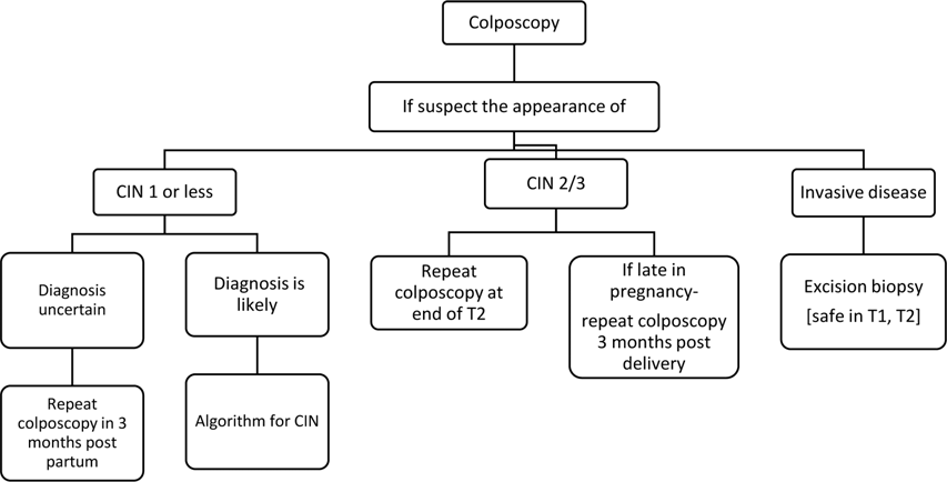
T1 - first trimester, T2 - second trimester
Flowchart 5: Pathway following the hysterectomy and CIN
Abbreviations
ASCUS Atypical Squamous Cells of Undetermined Significance
CA Cancer
CIN Cervical Intraepithelial Neoplasia
CKC Cold Knife Conization
HG High Grade
HIV Human Immunodeficiency Virus
HLD High Level Disinfection
HR-HPV High risk Human Papilloma Virus
HPV DNA Human Papilloma Virus Deoxyribo Nucleic Acid
HPV Human Papilloma Virus
HSIL High Grade Squamous Intraepithelial Lesion
LEEP Loop Electrosurgical Excision Procedure
LG Low Grade
LLETZ Large Loop Excision of the Transformation Zone
LSIL Low Grade Squamous Intraepithelial Lesion
NILM Negative for Intraepithelial Lesion or Malignancy
SCJ Squamocolumnar Junction
T1 first trimester
T2 second trimester
TZ Transformation Zone
WWC Well Women Clinics
Annexure 1
High risk groups for cervical screening
- Contact with multiple sexual partners or partners having multiple sex partners
- Smokers
- Age above 40 years and ever been sexually active
- Sexual debut at very young age
- Multiparity, especially at young age
- Low socio-economic status
- Not being screened for cervical cancer
- Patients with STD infections like chlamydia, gonorrhea, herpes.
- HIV patients
- Immunodeficiency
- Previously abnormal cervical smear: low grade or above
Annexure 2
Sample colposcopy proforma:
- Reason for referral
- Grade of cytological abnormality
- Whether the examination was adequate or inadequate – for the examination to be adequate, the entire cervix must be seen
- The presence or absence of vaginal and/or endocervical extension
- The colposcopic features of any lesion
- The colposcopic impression of lesion grade
- The type of transformation zone, ie type 1,2 or 3
- The site of any colposcopically directed biopsies.
Annexure 3
Obtaining a sample
Cytology based sampling (Conventional)
Samples are not to be taken while during the menstruation or active infection of the cervix. A cervical smear should be always be done prior to bimanual vaginal examination. Insert an appropriately sized Cusco’s speculum and inspect the cervix (warm water may be used to lubricate the speculum if necessary).
- A sample of cells is taken from the transformation zone of the cervix using a wooden spatula with an extended tip and a cytobrush.
- Ideally, two smears should be prepared on two glass slides.
- Each smear should be labeled clearly.
- The entire transformation zone should be sampled as almost all high-grade lesions develop in this zone.
- The smears should be immediately fixed in a 95% alcohol solution to preserve the cells.
- Fixed and air-dried slide is sent to a cytology laboratory.
- No smears should be repeated within three months as time is needed for the regeneration of the epithelium. Therefore, if a smear needs repeating, it should be done only after 3 months.
Liquid based cytology sampling
After visualization of the cervix as in above,
- Gently wipe away excessive discharge/mucous on the cervix with an oversized cotton swab.
This should be done as gently as possible to avoid removing the cervical cells to be sampled.
- Insert the broom deeply enough into the endocervical canal so that the shorter bristles fully contact the
ectocervix. Rotate the broom 5 times in a clockwise direction.
Alternatively, spatula can be inserted into the cervical os and rotate 360° with firm pressure or
the cytobrush into the cervical os no further than the end of the bristles and rotate 90°.
- Transferring the cells to the media
For SurePath: Insert the broom into the larger opening of the vial. Rotate the broom 90° to use the
inner edge of the insert to pull off the broom into the vial.
For ThinPrep: Rinse the broom by pushing it into the bottom of the vial 10 times, forcing the
bristles apart. Then, swirl the broom vigorously to release more material.
- Place the cap on the vial and tighten firmly.
References
1. Bruni L, Albero G, Serrano B, Mena M, Gómez D, Muñoz J, Bosch FX, de Sanjosé S. ICO/IARC Information Centre on HPV and Cancer (HPV Information Centre). Human papillomavirus and Related Diseases in Sri Lanka. Summary Report 17 June 2019. [Accessed on 13th May 2020]
2. Pesola F, Sasieni P. Impact of screening on cervical cancer incidence in England: a time trend analysis. BMJ Open 2019; 9: e026292. doi: 10.1136/ bmjopen-2018-026292
3. World Health Organization. Regional Office for South-East Asia. (2015). Strategic framework for the comprehensive control of cancer cervix in South-East Asia Region. World Health Organization, Regional Office for South-East Asia. https:// apps.who.int/iris/handle/10665/152098
4. National Colposcopy Training Module, Guideline and Standard Operating Procedure. National Cancer Control Programme, Ministry of Health and Indigenous Medical Services, Colombo, Sri Lanka; 2020.
5. Basu P, Sankaranarayanan R (2017). Atlas of Colposcopy – Principles and Practice: IARC CancerBase No. 13 [Internet]. Lyon, France: International Agency for Research on Cancer. Available from: https://screening.iarc.fr/atlascolpo. php, accessed on 30 June 2021.
6. Public Health England Guidance. NHS cervical screening (CSP) programme.Cervical screening: programme and colposcopy management. Guidance 4. Colposcopic diagnosis, treatment and follow up. Updated 5 February 2020 [Accessed on 13th May 2020]
7. Public Health England Guidance. NHS cervical screening (CSP) programme.Cervical screening: programme and colposcopy management. 8. Flowcharts: cervical screening protocol and colposcopy management recommendations. Updated 5 February 2020 [Accessed on 13th May 2020]
8. British Society for Colposcopy and Cervical Pathology (BSCCP) Cervical screening programme. Colposcopy and programme management. June 2020. Mobile application. [Accessed on 13th May 2020]
9. Guidelines for screening and treatment of precancerous lesions for cervical cancer prevention WHO guidelines, Publication date: 2013: ISBN: 978 92 4 154869 4
10. Sasieni P, Adams J. Effect of screening on cervical cancer mortality in England and Wales: analysis of trends with an age cohort model. BMJ 1999; 318(7193): 1244-5.
11. Sasieni P, Castanon A, Parkin DM. How many cervical cancers are prevented by treatment of screen-detected disease in young women? International Journal of Cancer 2009; 124(2): 461-4.
12. Saeed-Vafa, Huang Y. Should cervical cancer screening begin at 21 for everyone? A quantitative analysis in a high-risk, low-income, African American/Hispanic young-adult population. Diagnostic Cytopathology, 2013, forthcoming.
13. Rebolj M, van Ballegooijen M, Lynge E et al. Incidence of cervical cancer after several negative smear results by age 50: prospective observational study. BMJ 2009; 338: b1354.
14. Guidance on the use of liquid-based cytology for cervical screening (Technology Appraosal Guidance 69). London: NICE, 2003.
15. Moss S, Gray A, Legood R et al. Effect of testing for human papillomavirus as a triage during screening for cervical cancer: observational before and after study. BMJ 2006; 332: 83-5.
16. R S Kelly, J Patnick, H C Kitchener, and S M Moss, on behalf of the NHSCSP HPV Special Interest Group. HPV testing as a triage for borderline or mild dyskaryosis on cervical cytology: results from the Sentinel Sites study. Br J Cancer. 2011; 105(7): 983-8.